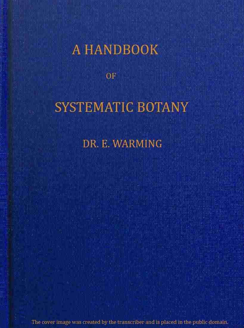
Fig. 85.—Empusa muscæ (Fly-mould). I. A fly killed by the fungus, surrounded by a white layer of conidia. II. The conidiophores (t) projecting from the body of the fly. Some of the conidia, a few of which have developed secondary conidia, are attached to the hairs (mag. 80 times). III. A perfect hypha. IV. A hypha in the act of ejecting a conidium (c), enveloped in a sticky slime (g). V. A conidium which has developed a secondary conidium (sc). VI. A branched hypha produced by cultivation. VII. A secondary conidium which has produced a small mycelium (m). VIII. A conidium germinating on the fly’s body. IX. Mycelium. X. Conidia germinating like yeast in the fatty tissue of the fly. (III.-VII. and IX. magnified 300 times; VIII. and X. magnified 500 times.)
A fertilisation, a passage of the contents of the antheridium to the egg-cell, has as yet only been observed in Pythium; in Phytophthora only one small mass of protoplasm passes through the fertilising tube to the egg-cell; in Peronospora and the Saprolegniaceæ no protoplasm can be observed to pass through the fertilising tube, so that in these instances parthenogenesis takes place; Saprolegnia thuretii, etc., have generally even no antheridia, but nevertheless form normal oospores. Fertilisation of the egg-cell by means of self-motile spermatozoids is only found in Monoblepharis sphærica.
A. Asexual reproduction by conidia only.
Family 1. Entomophthorales.
The mycelium is richly branched. The family is a transitional step to the conidia-bearing Zygomycetes, since the oospores of many members of this family arise, and are formed, like zygospores.
Order 1. Entomophthoraceæ. Mycelium abundantly developed. This most frequently lives parasitically in living insects, causing their death. The conidiophores forming the conidial-layer project from the skin, and abstrict a proportionately large conidium which is ejected with considerable force, and by this means transferred to other insects. These become infected by the entrance of the germ-tube into their bodies. The spherical, brown resting-spores develope inside the bodies of insects and germinate by emitting a germ-tube.
Genera: Empusa has a good many species which are parasitic on flies, moths, grasshoppers, plant-lice. The conidia emit a germ-tube which pierces the skin of the insect; a number of secondary conidia are then produced inside its body, by division or by gemmation similar to that taking place in yeast, each of which grows and becomes a long unbranched hypha, and these eventually fill up the body of the animal, causing distension and death. Each of these hyphæ projects through the skin, and abstricts a conidium, which is ejected by a squirting contrivance. The best known species is E. muscæ (Fig. 85), which makes its appearance epidemically towards autumn on the common house-fly, and shows itself by the dead flies which are found on the windows and walls attached by their probosces, distended wings, and legs. They have swollen abdomen, broad white belts of hyphæ between the abdominal rings, and are surrounded by a circle of whitish dust formed by the ejected conidia.—Entomophthora sends out, at definite places, from the mycelium hidden in the insect’s body, bundles of hyphæ, which serve the purpose of holding fast the dead insects, the ramifications attaching themselves to the substratum: the conidiophores are branched, the conidia are ejected by the divisional walls between the hyphæ and the conidia dividing into two layers, those which terminate the hyphæ suddenly expanding and throwing the conidia into the air. E. radicans makes its appearance epidemically on caterpillars.
B. Asexual reproduction by zoospores or conidia.
Family 2. Chytridiales.
In this family the mycelium is very sparsely developed or is wanting. The entire plant consists principally or entirely of a single zoosporangium whose zoospores have generally one cilium. The resting-spores arise either directly from the zoosporangium, which, instead of forming zoospores, surrounds itself by a thick cell-wall; or they are formed by the conjugation of two cells (in which case they are spoken of as oospores). Microscopic Fungi, parasitic on water plants (especially Algæ) or small aquatic animals, seldom on land plants.
Order 1. Olpidiaceæ. Without mycelium. Swarmspores and resting-spores.
In the Olpidieæ, the swarmspores, probably, most frequently form themselves into a plasmodium (naked mass of protoplasm) which may become a single zoosporangium or a resting sporangium. Olpidium trifolii occurs in Trifolium repens.—In the Synchytrieæ the plasmodium emerging from the swarmspores breaks up either at once, or after a period of rest, into smaller plasmodia, each of which will become a zoosporangium. Synchytrium anemones is found on Anemone nemorosa; S. mercurialis on Mercurialis perennis; S. aureum on many plants, particularly Lysimachia nummularia.
Fig. 86.—Chytridium lagenula. Zoosporangium a before, b after the liberation of the swarmspores.
Fig. 87.—Obelidium mucronatum: m mycelium; s swarmspores.
Order 2. Rhizidiaceæ. Mycelium present. Zoospores and resting-spores.
Chytridium (Fig. 86). Obelidium (Fig. 87) is bicellular; the one cell is the mycelium, the other the zoosporangium; found on insects. The species of Cladochytrium are intercellular parasites on marsh plants. Physoderma.
Order 3. Zygochytriaceæ. Mycelium present. Zoospores and oospores. The latter are the product of the conjugation of two cells (Fig. 88).
Polyphagus euglenæ on Euglena viridis. Urophlyctis pulposa on species of Chenopodium.
Family 3. Mycosiphonales.
The mycelium is bladder-like or branched. Zoospores. Sexual reproduction by oospores, which are produced in oogonia. The latter are fertilised, in some forms, by the antheridium.
Order 1. Ancylistaceæ. The entire bladder-like mycelium is used for the construction of zoosporangia, oogonia, or antheridia. Lagenedium is parasitic on Spirogyra, etc.
Order 2. Peronosporaceæ. Almost entirely parasites. The unicellular, often very long and abundantly branched mycelium lives in the intercellular spaces of living plants, especially in the green portions, and these are more or less destroyed and deformed in consequence. Special small branches (suction-organs, “haustoria”) are pushed into the cells in order to abstract nourishment from them. Both oospores and conidia germinate either immediately, or they develope into sporangia with swarmspores, having always two cilia. Only one oospore is formed in each oogonium; its contents (Fig. 89) divide into a centrally placed egg-cell and the “periplasm” surrounding it; this is of a paler colour and on the maturity of the oospore forms its thick, brown, external covering.
Fig. 88.—Polyphagus euglenæ. A with smooth, B with thorny oospores; m and f the two conjugating cells.
Fig. 89.—Peronospora alsinearum. Mycelium with egg-cell and antheridium.
Fig. 90.—Phytophthora infestans (strongly magnified). Cross section through a small portion of a Potato-leaf (the under side turned upwards): a the mycelium; b b two conidiophores projecting through a stoma; c conidia; e the spongy tissue of the leaf; g the epidermis.
The Potato-fungus (Phytophthora infestans) is of great interest. Its thallus winters in the Potato-tuber; other organs for passing the winter, such as oospores, are not known. When the tuber germinates, the Fungus-hyphæ penetrate the young shoot and keep pace with the aerial growth and development of the plant. The conidiophores emerge through the stomata, especially on the under side of the leaves; they branch like a tree (Fig. 90), and appear to the naked eye as a fine mould on the surface of the plant. The disease soon makes itself known by the brown colouring of those parts of the plant which are attacked, and by their withering. An ovoid conidium arises at first by the formation of a dividing wall at the apex of each branch of the conidiophore (Fig. 90 c c), and immediately underneath it another is formed, which pushes the first to one side, and so on. These conidia sometimes germinate directly, and form a mycelium, but most frequently their protoplasm divides into many small masses, each of which becomes a pear-shaped zoospore provided with two cilia (Fig. 91). Water is required for their germination, and when the ripe conidia are placed in a drop of water the swarm-cells are formed in the course of about five hours. They swarm about in rain and dewdrops in the Potato-fields, and are carried with the water to the Potato-plants and to the tubers in the soil. The wind also very easily conveys the conidia to healthy Potato-fields and infects them. The enormous quantity of conidia and swarm-cells that may be formed in the course of a summer explains the rapid spreading of the disease; and the preceding makes it clear why wet summers are favourable to its existence. When the swarm-cells germinate, they round off, and then surround themselves with a cell-wall which grows out into the germ-tube, and pierces through the epidermis of the host-plant (Fig. 92). Having entered the host, a new mycelium is formed. The potato disease, since 1845, has been rampant in Europe; it has, no doubt, been introduced from America, which, it must be remembered, is the home of the Potato-plant.
Fig. 91.—Phytophthora infestans: a-c conidia detached; in c the swarm-cells are leaving the mother-cell; d two free-swimming swarm-cells.
Fig. 92.—Phytophthora infestans. Cross section through a portion of a Potato-stalk. Two germinating conidia (a, b) piercing the epidermis, and the mycelium penetrating the cells.
The conidia exhibit various characters which are employed for the separation of the genera. Pythium is the most simple form. The contents of the terminally-formed conidia emerge as a spherical mass and divide into swarmspores. P. de Baryanum lives in the seedlings of many different Flowering-plants, which it completely destroys.—Phytophthora is distinguished by the circumstance that the sparsely-branched conidiophores bear, sympodially, chains of conidia. Besides the Potato-fungus (see above), Ph. fagi belongs to this group; it developes oospores very abundantly, and does great harm to seedlings of the Beech, Sycamore, and Pine trees.—Peronospora generally has conidiophores which are repeatedly forked, and bear a conidium on each of the most extreme ramifications. Many do great harm to their host-plants. P. viticola, on Vines, and P. nivea, on umbelliferous plants, have swarmspores, which are absent in the following species of this genus: P. sparsa, on Roses; P. gangliformis, on composites; P. alsinearum, on Stitchwort; P. parasitica, on cruciferous plants; P. viciæ, on Vetches and Peas; P. schachtii, on Beets; P. violacea, on the flowers of Scabiosa; P. radii, on the ray-florets of Matricaria.—Cystopus (Albugo) has the conidia developed in chains, which form a cohesive white layer underneath the epidermis of the host-plant. Cystopus candidus, on cruciferous plants, especially Shepherd’s Purse and Brassica; the germination commences on the cotyledons, and from this point the mycelium developes together with the host-plant; C. cubicus, on the leaves of Compositæ.
Fig. 93.—A fly overgrown with Saprolegnia.
Fig. 94.—Formation of swarmspores in a Saprolegnia: a germinating swarmspores.
Order 3. Saprolegniaceæ, Water-Fungi which live as saprophytes on organic remains lying in water, for instance, on dead flies (Fig. 93), worms, remains of plants; but they may also make their appearance on living animals, being frequently found, for example, on the young trout in rearing establishments.
Fig. 95.—Oogonium with two antheridia, Achlya racemosa.
The thallus is a single, long and branched cell. It has one portion which serves as root, and lives in the substratum, where it ramifies abundantly for the purpose of absorbing nourishment; and another portion projecting freely in the water, and sending out hyphæ on all sides (Fig. 93). The asexual reproduction takes place by swarmspores (Fig. 94), which are developed in large sporangia; these swarmspores generally possess two cilia, and on germination grow into new plants. The entire protoplasm in the oogonium is formed into one or more oospheres, without any surrounding “periplasm.” The oospheres may not be fertilised (p. 100), and then develope parthenogenetically.
Genera: Saprolegnia, whose swarmspores disperse immediately after having left the sporangium. S. ferax is the cause of a disease in fish (“Salmon disease”) and in the crayfish.—Achlya, whose swarmspores accumulate in a hollow ball before the mouth of the sporangium.—Leptomitus has strongly indented hyphæ, causing a “linked” appearance. L. lacteus is frequent in the waste matter from sugar factories.—Monoblepharis deviates from the others by the greater development of its fertilising process; the oosphere, situated in an open oogonium, becoming fertilised by self-motile spermatozoids, which are provided with a cilium at the posterior end.
Class 2. Mesomycetes.
The Mesomycetes are intermediate forms between the Phycomycetes and the Higher Fungi. In the vegetative organs, and in the multicellular hyphæ, they resemble the Higher Fungi; the methods of reproduction, however, show the characters of the Phycomycetes, namely sporangia and conidiophores of varying size and with varying number of spores; definite and typically formed asci and basidia are not present. Sexual reproduction is wanting. The Hemiasci are transitional between the Phycomycetes and the Ascomycetes, the Hemibasidii (Brand-Fungi) form the transition to the Basidiomycetes.
Sub-Class 1. Hemiasci.
The Hemiasci are Fungi with sporangia which, although resembling asci, yet have not, however, a definite form and a definite number of spores. Besides endospores, conidia, chlamydospores and oidia are found.
Order 1. Ascoideaceæ. Ascoidea rubescens forms irregular, reddish-brown masses in the sap issuing from felled Beeches. It has free sporangia, which resemble asci in their structure, in the development and ejection, and in the definite shape and size of the spores. The formation of the sporangia takes place when the nutriment is nearly exhausted, and resembles that of the conidia, since they are developed from the end of a hypha which enlarges, and the swelling becomes separated by a transverse wall. Within the sporangia numerous spores of a cap-like form are developed, which are set free through an opening at the apex. Sporangia are formed successively at the apex of the same hypha, the second commencing to develope as the first is dehiscing. Conidia and sporangia are not formed simultaneously; the former may be considered as closed sporangia.
Order 2. Protomycetaceæ. Protomyces pachydermus causes hard swellings on the stems and leaf-stalks of the Cichorieæ (Taraxacum, etc.). These swellings consist of chlamydospores (resting-spores), which germinate and become free, ascus-like sporangia, with numerous small spores. In nutritive solutions the chlamydospores form conidia with yeast-like buddings. P. macrosporus on Ægopodium, and other Umbelliferæ.
Order 3. Thelebolaceæ. Thelebolus stercoreus, is found on the dung of deer, hares, and rabbits, and has closed sporangia, which resemble asci in their shape and regular construction, and in the ejection of spores. The covering encloses only one sporangium, even where the sporangia arise close together.
This order, by reason of the covering of the sporangia, forms the transition from the Hemiasci to the Carpoasci, while the two first supply an intermediate step to the Exoasci.
Sub-Class 2. Hemibasidii, Brand-Fungi.
The Brand-Fungi (also known as Ustilagineæ) are Fungi with basidia-like conidiophores, which, however, have not yet advanced to a definite form or number of conidia. They are true parasites, whose mycelium spreads itself in the intercellular spaces of Flowering plants. The mycelium is colourless, quickly perishable, has transverse walls at some distance from each other (Fig. 96), and sends out haustoria into the cells of the host-plant.
Fig. 96.—Entyloma ranunculi. 1. Cross section of a portion of a leaf of Ficaria permeated by the mycelium; a bundle of hyphæ with conidia emerging from a stoma; in one of the cells are found four brand-spores. 2. A brand-spore developed in the middle of a hypha.
It most frequently happens that the germ-tube enters the host-plant at its most tender age, that is, during the germination of the seed; the mycelium then wanders about in the tissues of the shoot during its growth, until it reaches that part of the plant where the spores are to be formed. The spore-formation takes place in the same way in all those species whose brand-spores are developed in the floral parts of the host-plant. Many Brand-Fungi have, however, a more local occurrence, and the mycelium is restricted to a smaller area of the leaf or stem. Those organs of the host-plant in which the brand-spores are developed often become strongly hypertrophied. In perennial plants the mycelium winters very often in the rhizome.
Fig. 97.—Doassansia alismatis. 1. A fruit-body, formed by a covering of oblong hyphæ, which encloses a mass of brand-spores, and is embedded in the leaf-tissue of the host-plant; 20 times natural size. 2. A germinating brand-spore, 500 times natural size. 3. Three connected resting-spores, 400 times natural size. 4. Two conidia grown together, 600 times natural size.
The brand-spores are the winter resting-spores of the Brand-Fungi. They arise in the tissues of the host-plant, which is often destroyed, and become free through the rupture of the epidermis; they are thick-walled, generally brown or violet, and very often possess warts, spines, or reticulate markings. Fruit-bodies, that is enclosed organs of reproduction, are found in few genera (Sphacelotheca, Graphiola; Doassansia, Fig. 97). In Tolyposporium, Tuburcinia, Thecaphora (Fig. 102), etc., the brand-spores are united into a ball of spores. On germination the brand-spores behave as chlamydospores, namely, as the fundament of conidiophores, by emitting a short germ-tube, i.e. a conidiophore (“promycelium”). The Ustilaginaceæ (Fig. 99, 2) have a short transversely divided conidiophore, with laterally developed conidia (comp. the basidia of the Protobasidiomycetes). The conidiophores of the Tilletiaceæ are undivided (unicellular promycelia), and bear the conidia terminally, and so resemble the basidia of the Autobasidiomycetes.
Fig. 98.—Tuburcinia. 1. T. trientalis. Hyphæ, some of which bear conidia at the apex, forcing themselves out between the epidermal cells on the under side of the leaf; 320 times natural size. 2. T. trientalis. A ball of spores in which some of the individual brand-spores are about to germinate; 520 times natural size. 3. T. primulicola: various forms of conidia (500 times natural size).
In Tilletia, Entyloma, Neovossia, Tuburcinia, the brand-spores germinate and form basidia-like conidiophores with spindle-shaped conidia; their mycelium, on the other hand, produces later only single, sickle-shaped conidia, so that two kinds of conidia are found, as in a few Basidiomycetes. In some species, e.g. Ustilago hordei, the brand-spores only germinate vegetatively and form a mycelium. In nutritive solutions (solutions of dung, etc.) where they live as saprophytes, the brand-spores of many species emit germ-tubes, and on these, yeast-like conidia are produced by repeated budding, which grow into mycelia only when the nutritive solution is exhausted. These conidia have not the power of producing alcoholic fermentation. The very numerous conidia, which are found in the dung of herbivorous animals, are probably the yeast-conidia of Brand-Fungi. The brand-spores, which are eaten by animals with the grain and hay, pass into the dung and without doubt give rise to a very rich multiplication of yeast-conidia.
Fig. 99.—Ustilago. 1. Formation of brand-spores. 2. Germinating brand-spore of U. perennans. 3. Germinating brand-spore of U. cardui (after Brefeld). 4. U. filiformis. a A brand-spore with developed basidium; b another, with a conidium; c with two conidia; d with two conidia placed diametrically opposite to each other; e, detached conidia which are growing into hyphæ.
Fig. 100.—Tilletia tritici: a an ear of Wheat in which all the grains are attacked by Stinkbrand; b a blighted corn surrounded by the chaff; c a blighted corn grown together with a stamen; d the same cut across; e a brand-spore; f, g, h germinating brand-spores; i germinating conidia; j the mycelium; k-k brand-spore-forming mycelium-threads. (c-h magnified 400 times; i-k 300 times.)
The conidia (also called “sporidia”) of many species unite generally into an H-form (Figs. 97, 4; 100 h; 101, 4). This union in pairs does not, however, take place with a view to germination, there is no fusion of nuclei, and therefore in this “fusion” there is no sexual act.
Order 1. Ustilaginaceæ. Conidiophores with transverse walls and lateral conidia.—Ustilago (Fig. 99) generally developes its spores in the floral organs of its host-plant, the ovary or anthers, where they arise from hyphæ, and form a slimy mass which when mature becomes a black dust.
To this order belong U. avenæ, parasitic on Oats, U. hordei and U. nuda (U. jenseni), on Barley; these are the usual cause of “Smut” on cereals. U. hypodytes on straw of Elymus and Agropyrum. U. filiformis in the leaves of Glyceria. U. caricis transforms the fruits of various species of Carex into black, dusty balls. U. violacea developes its violet spore-powder in the anthers of the Caryophyllaceæ. U. tragopogonis, transforms entire inflorescences of Tragopogon into a black-violet mass. Among the largest are U. grandis, which causes the large swollen nodes in the stem of Phragmites, and the Maize Blight, U. maydis, which produces outgrowths about the size of a hand on the spadix of the Maize.
Order 2. Tilletiaceæ. Conidiophores undivided, generally several conidia arise at their apices.—Tilletia tritici, the Stinkbrand on Wheat (Fig. 100). The mycelium lives in Wheat-plants, producing its spores in the ovary after the whole interior of this body has been destroyed by the mycelium, with the exception of the external layer of the wall of the ovary, which remains essentially unaltered and encloses the closely packed, firm mass of spores (Fig. 100 d). The grains of Wheat thus attacked are shorter and thicker than the sound ones, and the ears show the presence of this Fungus by their erect position, and the wide separation of the chaff (Fig. 100 a). The unpleasant odour of the ovary prior to the ripening of the spores, has given the name “Stinkbrand,” and, in like manner, its hardness when it encloses the ripe spores, is the reason of its being also called “Stonebrand.” On account of this hardness, the diseased grains are readily harvested together with the healthy ones, which become infected by the spores at the threshing. T. lævis (T. fœtens) also occurs on Wheat and has smooth brand-spores.
Fig. 101.—Urocystis. 1, U. covalloides. A spore-ball, magnified 450 times. 2–4, U. anemones: 2–3, brand-spores which are about to germinate (magnified 450 times). 4, Conidia, the two in a state of fusion, a third with vacuoles and division-wall, magnified 500 times.
Entyloma (Fig. 96), a genus with numerous species, which appear in spots on the leaves of the host-plant, and Tuburcinia (Fig. 98), which makes its appearance on the Primulaceæ, produce white conidia-spots on the surface of the host-plant. The first-named has single spores, the latter has its spores closely massed together.—Urocystis (Fig. 101) has its spores surrounded by a number of small and lighter coloured barren spores. U. occulta, Rye-stem Blight, forms its spores in long streaks in the stems and leaves of the Rye, and does considerable damage. U. cepulæ on Onions. U. violæ forms large dark-violet swellings in the leaf-stalk and stems of Violets.—Thecaphora (Fig. 102) appears in seedlings of Convolvulus and Astragalus.
As a means of protection against the Smut-Fungi which make their appearance on the different cereals, a submersion of the grains in a solution of blue vitriol (½%) for twelve hours, or better still, submerging for five minutes in water heated to 53–55° C (Jensen’s method) is employed.
Fig. 102.—Thecaphora. 1, T. convolvuli, a ball of spores, one of the brand-spores has emitted a septate branched conidiophore (× 520). 2, T. affinis, a ball of spores (× 520).
Class 3. Mycomycetes, Higher Fungi.
The Mycomycetes are not entirely aquatic in habit; they have hyphæ with transverse walls, but no sexual reproductive organs. The asexual reproduction takes place in very different ways; by endospores (in asci), conidia, basidiospores, chlamydospores, and oidia. Swarmspores are never found.
Two chief methods of reproduction may be distinguished, and hence the class may be divided into two large sub-classes:—the Ascomycetes (with asci), and the Basidiomycetes (with basidia).
Sub-Class 1. Ascomycetes.
The main characteristic which distinguishes the Ascomycetes is the ascus; a name given to a sporangium of a definite shape and size, and containing a definite number of spores. The shape is generally club-like or spherical, the number of spores 8 (in some 2, 4, 16 or more), see Figs. 103, 105, 108, 110, 113, 116, 120, 121, 123, 129.
In the lowest forms, the Exoasci, the ascus springs directly from the mycelium without the formation of a fruit-body (i.e. ascocarp). In the higher forms, which contain many species, the Carpoasci, the asci are united and form ascocarps which may be more or less enclosed (angiocarpic, hemiangiocarpic, and probably gymnocarpic).
Fig. 103.—Endogenous formation of spores in Peziza confluens. In the youngest asci there is only one nucleus (b, e); this divides into two (f); and the division is repeated so that there are 4 nuclei in c and 8 in g. These surround themselves with protoplasm and a cell-wall (h, i). The protoplasm of the mother-cell is not entirely used up.
The hyphæ of the Mycelium in some remain free, in others they are felted together and form thick strands or flat, cushion-like bodies (compare in particular the stromata of the Pyrenomycetes). Some species form sclerotia (Figs. 116, 128).
Asexual reproduction by means of conidia is known in many species as the principal means of reproduction, and the one which affords the most rapid means of distribution. The conidia may be produced on conidiophores (Fig. 109), in conidial-layers (Fig. 122), and often in conidiocarps (pycnidia, Figs. 120 d, e; 123 a; 124 b.). These last occur partly as the so-called “spermogonia” (that is, pycnidia with microconidia). The conidiophores never approach the basidia.
In many species the ascospores germinate and form conidia immediately (Nectria cinnabarina, Sclerotinia, Taphrina, etc.), sometimes while they are still in the ascus and before their ejection (Taphrina, Fig. 105 a). In many instances the conidia by means of continued budding can, for a longer or shorter time, produce yeast-conidia, e.g. Taphrina. In many other cases the conidia arise from the germ-tubes of the ascospores, or at any part of the mycelium. The unripe asci of Taphrina, when placed in water, develop conidia at their apices. The Sclerotinia-species produce numerous conidia whose germination has never been observed. The formation of conidia and asci sometimes takes place on the same fruit-body. In Heterosphæria patella the conidia and asci are developed successively in the same fruit-body; in the ascocarps of Dermatea frangula and Sclerotinia sclerotiorum the formation of conidia may take place. The ascocarps frequently arise from the conidial-layers (Nectria cinnabarina, etc.). This relationship of the two forms of reproduction to each other may be explained by considering that both have descended phylogenetically from sporangia.
Sometimes chlamydospores and oidia also appear in the Ascomycetes; on germination, however, they do not, as in Protomyces, form sporangia, and on this account cannot be distinctly distinguished from conidia.
The asci are morphologically the highest form of reproduction and are always found at the close of the development of these Fungi; the accessory forms of reproduction are first developed, but a well-defined alternation of generations does not occur.
In the Ascomycetes there are more than 11,000 described species, which can be classed as follows:—
- Series 1. Exoasci. Only one order.
- „ 2. Carpoasci.
- Family 1. Gymnoascales, }
- „ 2. Perisporiales, } Angiocarpic Carpoasci.
- „ 3. Pyrenomycetes, }
- „ 4. Hysteriales, }
- „ 5. Discomycetes,} Hemiangiocarpic Carpoasci.
- „ 6. Helvellales, Gymnocarpic (?) Carpoasci.
- Additional Ascolichenes: Lichen-forming Ascomycetes.
Series 1. Exoasci.
Ascomycetes with FREE ASCI; sometimes also conidia, chlamydospores and oidia. One order.
Order. Taphrinaceæ. Of the genera belonging to this order, Taphrina, Endomyces, and Ascocorticium, the first is most important.
Endomyces decipiens is a parasite in the fruit-body of Armillaria mellea; E. magnusii lives in the gelatinous, fermenting exudations of Oak-trees; Ascocorticium albidum is found under the bark of the Fir-tree. Endomyces has chlamydospores and oidia.
The species of Taphrina are parasites, whose free asci may be found in great numbers, generally closely pressed together, on the parts of plants which they have attacked. The asci are developed directly from the ascogenous cells of a fertile, generally sub-cuticular, hypha, which arises from the sterile mycelium. The latter arises from the germinating ascospore, and may hibernate in the tissues of its host, particularly in the winter buds, and then with the commencement of the next period of vegetation it continues its growth side by side with that of its host. The hyphæ ramify in the intercellular spaces or beneath the cuticle, but have no haustoria. The ascospores (Fig. 105 A) and unripe asci may produce conidia.
Fig. 104.—Taphrina (Exoascus) pruni. Yeast-like budding of a germinating spore (× 600).
Fig. 105.—Taphrina betulina: a ascus filled with conidia; b germinating spores (× 600).
Very remarkable appearances, and swellings of the attacked tissues, are produced when the mycelium is perennial; for example, the “Witches’-brooms” and “Pockets.” The hard, hollow, stoneless plums, known as “Pocket” or “Bladder” Plums, are produced by considerable changes in the tissues of the fruit; these are caused particularly by T. pruni on several species of Prunus. The “Witches’-brooms,” on the contrary, are deformations of entire twigs or branches, and often attain a very large size. They occur on Alnus incana, caused by T. epiphylla; on Carpinus betulus, by T. carpini; on Cherry-trees, by T. cerasi; on Plum-trees, by T. insititiæ; on Birches, by T. turgida and T. betulina. T. deformans attacks the leaves of the Peach, and causes them to curl.
When a perennial mycelium is wanting, the infection is confined as a rule to white or yellow spots on the leaves, e.g. the commonest, T. sadebeckii, on Alnus glutinosa, and T. aurea on species of Populus. T. alni incanæ (Fig. 106) causes considerable hypertrophies on the pistillate catkins of the Alder, which may be compared to the “pockets” of Prunus.
Fig. 106.—Taphrina alni incanæ on the Alder (nat. size).
Series 2. Carpoasci.
The Carpoasci are Ascomycetes, whose asci are enclosed in fruit-bodies, i.e. ascocarps. The accessory means of reproduction are free conidiophores (Fig. 109), conidial-layers (Fig. 122), conidiocarps (Fig. 120 D, E, etc.), chlamydospores and oidia.
For the different methods of distributing the ascospores, see p. 92.
Of the six families of the Carpoasci, the first three—Gymnoascales, Perisporiales, and Pyrenomycetes—are ANGIOCARPIC (that is, the ascocarp remains closed throughout its existence, and does not dehisce when ripe); the fourth and fifth families (Hysteriales and Discomycetes), on the other hand, are HEMIANGIOCARPIC (the ascocarp, here also called an apothecium, is closed in the early stages, but opens at the commencement of ripening and exposes a hymenium of crowded asci); the family of Helvellales has probably GYMNOCARPIC (or hemiangiocarpic) fruit-bodies.
Family 1. Gymnoascales.
The ascocarps are surrounded by a spongy and incomplete envelope. One order, poor in species.
Order Gymnoascaceæ.—The ascocarps are borne sometimes solitarily, or sometimes coiled together. Gymnoascus reessii forms small bodies about 1 mm. in diameter on old horse-dung, which at first are white and afterwards orange-red.—Ctenomyces serratus lives on the old feathers in birds’ nests.
Family 2. Perisporiales.
The ascocarps are surrounded by a complete envelope without any opening: the fruit-bodies are cleistocarpic; the spores are only liberated after the disintegration of the fruit-bodies. Paraphyses are wanting. The two first orders have in addition the means of reproduction by conidia.
Order 1. Erysiphaceæ, Mildews. The Fungi belonging to this order are epiphytic parasites, whose mycelium, somewhat resembling a cobweb, may be seen on the leaves and other green portions of plants (see Figs. 107, 108). The hyphæ ramify in all directions upon the surface of their host, and emit haustoria which penetrate the epidermal cells, and thus derive the necessary nutriment. The Mildew-Fungi thus belong to the obligate parasites, and during their growth dwarf and destroy the portions of their host on which they live. The reproduction takes place in the first instance by abstriction of conidio-chains from the end of special branches (Fig. 108 c, a hypha is seen in the act of detaching a conidium). The conidia may germinate immediately, and thus quickly reproduce their species. When present in large numbers they appear as a white meal covering the surface of the plant on which the fungus is found. Later on appear the dark brown, spheroid ascocarps (Fig. 108 a) which, although small, are generally just visible to the naked eye as black specks.