Fig. 227.—View looking down upon the upper (i.e. exposed) surface of one of the large scales situated on the veins of Danais plexippus, about four days before emergence: clm, chitinous pillars found in scales. A, a smaller scale, a, a′, sections of the scales. B, leucocyte found in the larger scale.—After Mayer.
Fig. 228.—Scale-follicles: A, of a scale of Galleria mellonella: r, neck-ring. B, the same of Polyommatus phlæas. C, the same of a hair on inner edge of hind wing of Lycæna alexis ♀.—After Spuler.
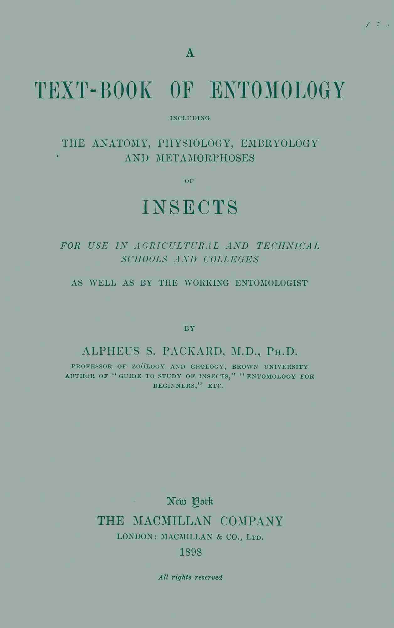
Fig. 229.—A, portion of wing of a caddis-fly (Mystacides). B, enlarged, showing the androconia and hair-scales. C, a separate androconium.—After Kellogg.
The “hair-scales” of the phylogenetically older Trichoptera correspond to certain scales of Lepidoptera, especially the Psychidæ (Spuler), variously called “plumules” (Deschamps), “battledore scales,” also certain minute cylindrical hairs. To these scent-scales is applied the term androconia. They are found, almost without exception, on the upper side of the fore wings, occurring in limited areas, such as the discal spots, or on folds of the wings. Fritz Müller has shown that they function as scent-scales, and are confined to the males. Kellogg has detected androconia-like scales on the wings of a caddis-fly, Mystacides punctata (Fig. 229).
Fig. 280.—Cross-section of androconia surface on wing of Thecla calanus; a, androconia; gl, gland of base; s, ordinary scales; w, wing in section.—After Thomas.
Thomas has proved by sections of the wing of Danais, etc., that the androconia arise from glands situated in a fold of the wing (Fig. 230), and he states that the material elaborated by the local glands, and distributed upon the surface of the wing by the androconia, is that which gives to many of the Lepidoptera their characteristic odor. On comparing these “glands,” it is evident that they are groups of specialized formative cells of Semper (trichogens), which secrete an odorous fluid, issuing perhaps from extremely fine pore-canals at the ends of the androconia. They thus correspond to the glandular hairs, poison-hairs, and spines of caterpillars, the formative cells of which contain either a clear lymph or poison.
LITERATURE
a. Hairs, bristles, cleaning spines, calcaria, combs, etc.
Leydig, Franz. Zum feineren Bau der Arthropoden. (Müller’s Archiv f. Anat. und Phys., 1855, pp. 376–480.)
Fobel, Auguste. Les fourmis de la Suisse. Bâle, 1874.
Saunders, Edward. Remarks on the hairs of some of our British Hymenoptera. (Trans. Ent. Soc. London, 1878, pp. 169–171.)
Perez, J. Notes d’apiculture. (Bull. Soc. d’Apic. de la Gironde, Bordeaux, 1882.)
Osten Sacken, C. R. von. An essay on comparative chætotaxy, or the arrangement of characteristic bristles of Diptera. (Trans. Ent. Soc. London, 1884, pp. 497–517.)
—— Preliminary notice of a subdivision of the suborder Orthorrhapha Brachycera (Diptera) on chætotactic principles. (Berlin Ent. Zeitschr., 1896, pp. 365–373.)
Janet, Charles. Études sur les fourmis. 8e Note. Sur l’organe de nettoyage tibio-tarsien de Myrmica rubra. (Ann. Soc. Ent. France, 1895, pp. 691–704, 6 Figs.)
See also J. B. Smith’s Economic Entomology, 1896, hairs of bees. Also the writings of De Geer, Huber, Fenger, Mayr, Forel, Canestrini, and Berlese (1880); Dahl, Cheshire, etc.
b. Glandular and poisonous setæ and spines
Ratzeburg, J. Th. Ch. Ueber entomologische Krankheiten. (Stettin Ent. Zeit., 1846, vii, pp. 35–41.)
Zeller, P. C. Revision der Pterophoriden. (Linnæa Ent., vi, pp. 319–416, 1852, at p. 356 speaks of “Drüsenhärchen.”)
Dimmock, George. On some glands which open externally on insects. (Psyche, iii, pp. 387–401, 1882.)
Goossens, Th. Des chenilles urticants. (Ann. Soc. Ent. France, 1881, pp. 231–236.) Des chenilles vésicants. (Ibid., 1886, pp. 461–464.)
Packard, A. S. Notes on some points in the external structure and phylogeny of insects. (Proc. Boston Soc. Nat. Hist., xxv, 1890, pp. 83–114, 2 Pls.)
—— A study of the transformations and anatomy of Lagoa crispata, a bombycine moth. (Proc. Amer. Phil. Soc., xxxii, pp. 275–292, 7 Pls., 1894.)
Holmgren, Emil. Studier öfner hudens och de körtelartade hudorganens morfologi hos Skandinaviska macrolepidopterlarver. (K. Svenska Vetenskaps-Akad. Handl., xxvii, pp. 1–83, Stockholm, 1895, 9 Pls.)
Also the writings of Leydig, Keller, Bach, Karsten, Scribner, Riley, etc.
(See also Literature of repugnatorial glands.)
c. Androconia
Deschamps, Bernard. Récherches microscopiques sur l’organisation des ailes der Lépidoptères. (Ann. des Sc. nat. [?], iii, pp. 111–157, 1835.)
Waufor, T. W. On certain butterfly scales characteristic of sex. (London, 1867–68.)
McIntire, S. J. Notes on the minute structure of the scales of certain insects. (London, 1871.)
Anthony, J. The markings on the battledore scales of some of the Lepidoptera. (London, 1872.)
Scudder, S. H. Antigeny or sexual dimorphism in butterflies. (Proc. Amer. Acad. Arts and Sc., xii, 1877, pp. 150–158.) Also Butterflies, etc. (New York, 1881, pp. 192–206, figs.)
Müller, Fritz. A prega costal das Hesperideas. (Archivas do Museo nac. do Rio de Janeiro, iii, pp. 41–50, 2 Pls., 1878.)
Thomas, M. B. The androconia of Lepidoptera. (Amer. Nat., xxvii, pp. 1018–1021, 2 Pls., 1893.)
d. Scales
Leydig, Franz. Zum feineren Bau der Arthropoden. (Archiv f. Anat. und Phys., 1855, pp. 376–480, 1 Taf.)
Semper, Carl. Beobachtungen über die Bildung der Flügel, Schuppen, und Haare bei den Lepidopteren. (Zeitschrift f. wissensch. Zoologie, 1857, pp. 326–339, 1 Taf.)
Mayer, F. T. Karl. Ueber den Staub der Schmetterlingsflügel. (Allgem. mediz. Centralzeitung, 1860, pp. 772–774.)
Landois, H. Beiträge zur Entwicklungsgeschichte der Schmetterlingsflügel in der Raupe und Puppe. (Zeitschr. f. wissensch. Zoologie, xxi, 1871, pp. 305–316, 1 Taf.)
Weismann, August. Ueber Duftschuppen. (Zool. Anzeiger, i, 1878, pp. 98–99.)
Dimmock, George. Scales of Coleoptera. (Psyche, iv, pp. 1–11, 23–27, 43–47, 63–71, 1883.)
Schaeffer, Cäsar. Beiträge zur Histologie der Insekten. (Zool. Jahrbücher, Abth. f. Anat. u. Ontog., iii, pp. 611–652, 2 Pls., 1889.)
Kellogg, Vernon L. The taxonomic value of the scales of the Lepidoptera. (Kansas Univ. Quart., iii, pp. 45–89, figs. 1–17, 9 Taf., 1894.)
Mayer, Alfred G. The development of the wing-scales and their pigment in butterflies and moths. (Bull. Mus. Comp. Zool., xxix., 1896, pp. 209–236, 7 Pls.)
Spuler, Arnold. Beiträge zur Kenntniss des feineren Baues und der Phylogenie der Flügeltedeckung der Schmetterlings. (Zool. Jahrb. Abth. f. Anat. u. Ontog., viii, pp. 520–543, 1 Taf., 1895.)
—— Ueber das Vorhandensein von Schuppenbalg bei den Schmetterlingen. (Biol. Centralblatt, xvi, Sept. 15, 1896, pp. 677–679, 3 figs.)
THE COLORS OF INSECTS
The colors and bright markings of insects, especially those of butterflies, render them the most brilliant and beautiful creatures in existence, rivalling and even excelling the gay hues of our most splendidly colored birds. The subject has been but recently taken up and is in a somewhat crude condition, but the leading features have been roughly sketched out by the work of a few observers from a physical, chemical, and biological point of view.
The colors of insects, as of all other animals, are primarily due to the action of light and air; other factors are, as Hagen observes, heat and cold, moisture and dryness, as recently shown by the experiments on butterflies by Dorfmeister, Weismann, W. H. Edwards, and later observers. They have their seat in the integument. Hagen divides colors into optical and natural.
Optical colors.—“These,” says Hagen, “are produced by the interference of light, and are by no means rare among insects, but they are solely optical phenomena. Colors by the interference of light are produced in two different ways: either by thin superposed lamellæ, or by many very fine lines or small impressions in very close juxtaposition.
“1. There must be present at least two superposed lamellæ to produce colors by interference. The naked wings of Diptera, of dragon-flies, and of certain Neuroptera often show beautiful interference colors. The wings of Chrysopa and Agrion show interference colors only for a certain time, viz., as long as the membranes of the wings are soft and not firmly glued together. Afterwards such wings become simply hyaline.
“The scales of Entimus and other Curculionidæ are well known for their brilliancy, and it is interesting to remark that when dry scales are examined with the microscope, many are found partly injured, which give in different places different colors, according to the number of layers which remain. The elytra of some Chrysomelina and other beetles with iridescent colors probably belong to the same category.
“2. When there are scales with many fine lines or small impressions close to each other, we have the second mode of producing colors.
“The fine longitudinal and transversal lines of lepidopterous scales seem to serve admirably well to produce the brilliant effect of color-changing butterflies. But there must be something more present, as most of the scales of Lepidoptera are provided with similarly fine lines, and only comparatively few species change colors. I remark purposely that the lines in the color-changing scales are not in nearer juxtaposition.” (Hagen.)
“The colors of butterflies change mostly from purple to blue, sometimes to yellow. The splendid violet color at the end of the wings of Callosune ione is brought out by a combination of the natural with interference colors. Originally the scales are colored lake-red; but a blue interference color is mixed with it; hence the violet hue results. The blue tones, i.e. the splendid varying blue of the Morpho butterflies, Schatz claims, owe their hue less to the interference of light than to a clouded layer of scales situated over the dark ground, through which the light becomes reflected on the same. The scales of the Morphids are in reality brown, as we see by transmitted light; moreover, only the upper side of the scales sends off blue reflections—the under side is simply brown. But the blue scales of Urvilliana are also shining blue beneath; by transmitted light they appear as if clear yellow. The smaragd-green scales of Priamus show by transmitted light a bright red-orange, and the orange-yellow of Crœsus a deep grass-green.” (Schatz in Kolbe.)
“Krukenberg presumes the golden-green color of Carabus auratus to be an interference color. It is not changed by the interference of light, nor was he able to extract from the elytra any green pigment with ether, benzol, carbon of sulphur, chloroform, or alcohol, even after having previously submitted the elytra to the influence of muriatic acid or ammonia. Chlorophyll is not present, whether free or combined with an acid.” (Hagen.)
Leydig has shown that the interference colors of the hairs of certain worms (Aphrodite and Eunice) may be produced by very small impressions in juxtaposition, which bring about the same effect as striæ. Such an arrangement occurs on the feathers of birds, i.e. on the necks of pigeons and elsewhere, and Hagen suggests that this kind of interference colors occurs more frequently among insects than is commonly known. At least the limbs of certain forms appear yellow, but when held in a certain position change to brown or blackish. “I know of no other explanation of this not uncommon fact on the legs of Diptera, of Hymenoptera, and of Phryganidæ.” Interference colors, he adds, may occur in the same place together with natural colors. “The mirror spots of Saturnia pernyi show besides the interference colors a white substance in the cells of the matrix, which Leydig believes to be guanin. But this fact is denied by Krukenberg for the same species and also for Attacus mylitta and Plusia chrysitis.”
Natural colors.—These are divided by Hagen into dermal (cuticular) and hypodermal. The dermal colors are due to pigment deposited in the form of very small nuclei in the cuticula. Hagen considers them as “produced mostly by oxidation or carbonization, in consequence of a chemical process originating and accompanying the development and the transformations of insects.”
“To a certain extent the dermal colors may have been derived from hypodermal colors, as the cuticula is secreted by the hypodermis, and the colors may have been changed by oxidation and air-tight seclusion. The cuticula is in certain cases entirely colorless,—so in the green caterpillar of Sphinx ocellata; but the intensely red and black spots of the caterpillar of Papilio machaon belong to the cuticula, and only the main yellow color of the body to the hypodermis.” (Leydig, Histiol., p. 114.)
“The dermal colors are red, brown, black, and all intermediate shades, and all metallic colors, blue, green, bronze, copper, silver, and gold. The dermal colors are easily to be recognized as such, because they are persistent, never becoming obliterated or changed after death.” (Hagen.)
Minot and Burgess refer to the cuticular colors of the cotton-worm (Aletia), the dark brown color belonging to the cuticula or crust. “Upon the outside of the crust is a very thin but distinct layer, which in certain parts rises up into a great number of minute, pointed spines that look like so many dots in a surface view. Each spine is pigmented diffusely, and together they produce the brown markings. The spines are clustered in little groups, one group over each underlying hypodermal cell.” (U. S. Ent. Comm., 4th Report, p. 46.) Minot also shows that in caterpillars generally a part of the coloration is caused by pigmentation of the cuticula.
In a dull-colored insect, such as the Mormon cricket (Anabrus), the coloration, as Minot states, depends principally upon the pigment of the hypodermis shining through the cuticula. “Most of the cells contain dull, reddish-brown granules, but scattered in among them are patches of cells bright green in color. I have observed no cells intermediate in color; on the contrary, the passage is abrupt, a brown or red cell lying next a green one. Indeed, I have never seen any microscopic object more bizarre than a piece of the epidermis of Anabrus spread out and viewed from the surface.” (2d Report U. S. Ent. Comm., p. 189.)
The pigment may extend through the entire cuticula, but it is usually confined to the outermost layers, and occurs there in union with a peculiar modelling of the upper surface into microscopic figures which are of interest not only from their delicacy, but because they vary with each species. (See p. 184.)
The hypodermal colors, situated in the hypodermis, are, according to Hagen, the result of a chemical process, generating color out of substances contained in the body. They are easily recognized, since they fade, change, and disappear after death. But where these colors are preserved after death and enclosed in air-tight sacs, as in the elytra and scales and hairs of the body, they persist, though, as we well know, they may fade after exposure to light.
The hypodermal colors are mostly brighter and lighter than the dermal ones, being light blue or green in different shades, yellow to orange, and the numerous shades of these colors combined with white; exceptionally they are metallic, as in Cassida, and are then obliterated after death.
“The fact that such metallic colors can be retained in dead specimens by putting a drop of glycerine under the elytra, leads us to conclude that those colors are based upon fat substances. The hypodermal colors are never glossy, as far as I know; the dermal colors frequently.
“As the wings, elytra, and hairs all possess a cuticula, dermal colors are frequently to be found, together with hypodermal ones, chiefly in metallic colors. In the same place both colors may be present, or one of them alone. So we find hypodermal colors in the elytra of Lampyridæ. In the elytra of the Cicindelidæ the main metallic color is dermal, the white lines or spots are hypodermal, by which arrangement the variability in size and shape of those spots is explained.
“There occur in a number of insects external colors, that is, colors upon the cuticula, which I consider to be in fact displaced hypodermal colors: the mealy pale blue or white upon the abdomen of some Odonata, the white on many Hemiptera, the pale gray on the elytra and on the thorax of the Goliath beetle, and the yellowish powder on Lixus. Some of these colors dissolve easily by ether or melt in heat, and some of them are a kind of wax. I believe that those colors are produced in the hypodermis, and are exuded through the pore-canals.” (Hagen.)
The white colors are simply for the most part due to the inclusion of air in scales. The white mother-of-pearl spots of Argynnis are produced by a system of fine transverse pore-canals filled with air; in Hydrometra the white ventral marks have the same origin. (Leydig.)
The further statements and criticisms of Hagen regarding the relation of color to mimicry, sexual selection, and the origin of patterns are of much weight and will be referred to under those heads. Indeed, these subjects cannot well be discussed without reference to the fundamental facts stated in the masterly papers of Leydig and of Hagen, and much of the theorizing of these latter days is ill-founded, because the colors of insects and animals are attributed to natural selection, when they seem really the result of the action of the primary factors of organic evolution, such as changes of light, heat, cold, and chemical processes dependent on the former.
As to the chemical nature of color, Hagen, after quoting the results of Krukenberg and others, thinks that the colors of insects are chemically produced by a combination of fats or fat-acids with other acids or alkalis under the influence of air, light, and heat. He concludes:—
1. That some colors of insects can be changed or obliterated by acids.
2. That two natural colors, madder-lake and indigo, can be produced artificially by the influence of acid on fat-bodies.
3. As protein bodies in insects are changed into fat-bodies, and may be changed by acids contained in insects into fat-acids, the formation of colors in the same manner seems probable.
4. That colors can be changed by different temperatures.
5. That the pattern is originated probably by a combination of oxygen with the integument.
6. That mimicry of the hypodermal colors may be effected by a kind of photographic process.
7. Finally, color and pattern are produced by physiological processes in the interior of the bodies of insects.
Krukenberg concludes that change of color (in perfectly developed insects) is a consequence of the change of food, and can be explained by the alteration of the pigment through heat and light. His experiments were made in order to ascertain the cause of the turning of green grasshoppers in autumn into yellow and pink. He tried to answer two questions: First, does the pigment of grasshoppers originate directly out of the food, and does it consist of pure chlorophyll or of a substance containing chlorophyll, or is it to be accepted as a peculiar product of the organism? Second, is the color the consequence of only one pigment, or of several? Special analysis proves that the green color has no connection with chlorophyll. He concludes: “It is evident that the green color of the grasshopper is the consequence of several different pigments which can be separated by a chemical process.” Krukenberg believes that light has a marked influence on the color of insects and that light turns to red or pink the insects which were green during the summer. It would seem, however, more probable that cold was the agent, the change being due to the colder autumn weather.
Here we might refer to the results of the studies of Buckton and Sorby, on the changes in color of Aphides:—
“1. The purple coloring matter appears to be a quasi-living principle, and not a product of a subsequent chemical oxidizing process. Mounted in balsam or other preserving fluids, the darker species stain the fluid a fine violet.
“2. As autumn approaches and cold weather reduces the activity of the Aphides, the lively greens and yellows commonly become converted into ferruginous red, and even dark brown, which last hue in reality partakes more or less of intense violet or purple. These changes have some analogy with the brilliant hues assumed by maple and other leaves during the process of slow decay.
“3. Aqueous solutions of crushed dark brown and yellow-green varieties of Aphides originate different colors with acids and alkalies.
“4. In the generality of cases coloring-matters, such as indigo, Indian yellow, madder-lake, and the like, do not separately exist in the substance of vegetables, but the pigments are disengaged through fermentation or oxygenation. Again, alizarin itself is reddish yellow, but alkaline solutions strike it a rich violet just as we find them to act towards the substance which Mr. Sorby calls aphidilutein.
“5. Mr. Sorby’s four stages of the changes effected by the oxidation of aphideine produce four different substances.”
Chemical and physical nature of the pigment.—Researches in this difficult field of inquiry have been made by Landois (1864), Sorby (1871), Meldola (1871), by Krukenberg (1884), and more recently by Coste, Urech, Hopkins, and Mayer, and the subject is of fundamental importance in dealing with mimicry and protective coloration, the primary causes of which appear to be due to the action of physical and chemical agents.
Over twenty years ago Meldola observed that the yellow pigment of the sulphur-yellow butterfly (Gonopteryx rhamni) was soluble in water, and showed that its aqueous solution had an acid reaction.
Besides the yellow uranidin found by Krukenberg in different beetles and lepidopterous pupæ, still other coloring-matters, which are very constant in different species are readily recognized by the spectroscope. “Thus there appear in the brownish yellow lymph of Attacus pernyi, Callosamia promethea and Telea polyphemus, after saponification of the precipitated soap readily effected by ether, or incompletely or not removed by benzine, a chlorophane-like lipochrome; and in the yellowish green lymph of Saturnia pyri and of Platysamia cecropia besides this pigment still another whose spectrum shows a broad band on D, but which disappears with the addition of acetic acid or ammonia, as also after a long heating of the lymph up to 66° C.”
Coste, and more especially Urech, have shown that many of the pigments may be dissolved out of the scales by means of chemical reagents, giving colored solutions, and leaving the scales white or colorless. They have also shown that some of these pigments may be changed in color by the action of reagents, and then restored to their original color by other reagents. They have proved that reds, yellows, browns, and blacks are always due to pigments, and in a few cases greens, blues, violets, purples, and whites, and not, as is usually the case, to structural conditions, such as striæ on the scales (Mayer). They confined themselves solely to the chemical side of the problem, not considering the structure of the scales themselves.
Urech has also discovered a beautiful smaragd-green coloring-matter in the wings (not in the scales) of the pupa of Pieris brassicæ. It is not chlorophyll, and Urech suggests that it may be either the germinal substance of the pigments of the scales or its bearer. It is not the pigment of the blood.
Urech has also demonstrated that in many Lepidoptera the color of the urine which is voided upon emergence from the chrysalis is similar to the principal color of the scales.
Hopkins has worked on the pigments within the scales of butterflies. The yellow pigment in Gonopteryx rhamni is a derivation of uric acid, and he calls it lepidotic acid. Its aqueous solution is strongly acid to litmus, and must be bad-tasting to birds.
Hopkins has dissolved the red pigment from the border of the hind wing of Delias eucharis, an Indian butterfly, in pure water, finding as the result a yellow solution; but if the solution be evaporated to dryness, the solid residue of pigment is red once more. He has obtained from this pigment of eucharis a silver compound which contains a percentage of metals exactly equal to that from the pigment of G. rhamni. (Nature, April 2, 1892.)
“The scales of the wings of the white butterflies (Pieridæ) are also shown by Hopkins to contain uric acid, this substance practically acting as a white pigment in these insects. A yellow pigment, widely distributed in the same family, is shown to be a derivative of uric acid, and its artificial production as a by-product of the hydrolysis of uric acid is demonstrated. That this yellow pigment is an ordinary excretory product of the butterfly is indicated by the fact that an identical substance is voided from the rectum on emergence from the pupa. These excretory pigments, which have well-marked reactions, are apparently confined to the Pieridæ, and are not found in other Rhopalocera. This fact shows that when a Pierid mimics an insect belonging to another group, the pigments of the mimicked and mimicking insects, respectively, are chemically quite distinct. Other pigments existing, not in the scales, but between the wing-membranes, are shown to be of use for ornament.” (Proc. Royal Soc., London, 1894.)
Griffiths (1892) claims that the green pigment found in several species of Papilio, Hesperia, and Limenitis, also in Noctuidæ, Geometridæ, and Sphingidæ likewise consists of a derivative of uric acid, which he calls lepidopteric acid. By prolonged boiling in HCl it is converted into uric acid.
Spuler, however, finds that green does not depend on pigmentation, but is an optical color. As remarked by Spuler, either the chitin of the scales itself is colored reddish (yellow grayish), or the pigment is secreted in the nuclei.
A. G. Mayer believes that the pigments of the scales are derived from the hæmolymph or blood of the pupa, for the following reasons: (1) He is unable to find anything but blood within the scales during the time when the pigment is formed. (2) In Lepidoptera generally the first color to appear upon the pupal wings is a dull ochre-yellow, or drab, and this is also the color assumed by the blood when it is removed from the pupa and exposed to the air. (3) He has succeeded by artificial means in manufacturing several pigments from the blood which are similar in color to various markings upon the wing of the imago; chemical reagents have the same effect upon these manufactured pigments that they do upon the similarly colored pigments of the wings. “It should be here noted,” he says, “that in 1866 Landois pointed out the fact that the color of the dried blood of many caterpillars is similar to the ground color of the wings of the mature insect.”
Ontogenetic and phylogenetic development of colors.—The colors of the wings of Lepidoptera, as is well known, are acquired at the end of the pupal state. The order of development of the colors in the pupal wings has been observed by Schaeffer, Van Bemmelen, Urech, Haase, Dixey, Spuler, and A. G. Mayer. The immature wings are at first transparent and full of protoplasm. The transparent condition of the wings corresponds to the period before the scales are formed, and when they are full of protoplasm; they then become whitish as the scales develop; the latter are at first filled with protoplasm, and afterwards turn whitish, being little hollow sacks filled with air. After the protoplasm has completely withdrawn from the scales, the blood of the pupa enters them, and then the coloring-matter forms. (Mayer.) He adds that “about twenty-four hours after the appearance of the dull yellow suffusion the mature colors begin to show themselves. They arise, faint at first, in places near the centre of the wings, and are distinguished by the fact that they first appear upon areas between the nervures, never upon the nervures themselves. Indeed, the last place to acquire the mature coloration are the outer and costal edges of the wings, and the nervures.”
The faint color of the scales gradually increases in intensity. “For example, if a scale be destined to become black, it first becomes pale grayish brown, and this color gradually deepens into black.”
Urech states that in Vanessa io first a white, and in V. urticæ a pale reddish hue, are spread over the entire wings, and then successively arise other colors in the following order: yellow, yellow to brown, red, brown and black.
Spuler, however, claims that the differentiation of colors and markings do not follow one another, but arise simultaneously, and that his view is confirmed by Fischer. This may be the case with the highly specialized and diversely marked butterflies, but certainly taking the Lepidoptera as a whole the yellows and drabs must have been the primitive hues, the other colors being gradually added in the later more specialized forms.
It is noticeable that the most generalized moths, such as the species of Micropteryx, Tinea, Psychidæ, Hepialidæ (in general), etc., are dull brown or yellow-drab without bars, stripes, or spots of bright hues. These shades prevail in others of the more primitive Lepidoptera, such as many bombycine moths, and they even appear to a slight extent in certain caddis-flies. The authors mentioned, especially Mayer, whom we quote, claim that “dull ochre-yellows and drabs are, phylogenetically speaking, the oldest pigmental colors in the Lepidoptera; for these are the colors that are assumed by the hæmolymph upon mere exposure to the air. The more brilliant pigmental colors, such as bright yellow, reds, greens, etc., are derived by more complex chemical processes. We find that dull ochre-yellow and drabs are at the present day the prevalent colors among the less differentiated nocturnal moths. The diurnal forms of Lepidoptera have almost a monopoly of the brilliant colorations, but even in these diurnal forms one finds that dull yellow or drab colors are still quite common upon those parts of their wings that are hidden from view.”
The more primitive moths being more or less uniformly yellowish or drab, the next step was the formation of bars, stripes, finally spots, and eyed spots, these markings in the later forms appearing simultaneously in one and the same species of certain highly specialized moths and butterflies. All that has been said will prepare the reader for the consideration of the subject of insect coloration. The origin of such markings has been discussed by Weismann, Eimer, Haase, Dixey, Fischer, and others.
LITERATURE
Heer, O. Einfluss des Alpenklimas auf die Farbe der Insecten. (Froebel u. Heer, Mitth. aus dem Gebiete der theoret. Erdkunde, 1836, i, pp. 161–170.)
Goureau. Mémoire sur l’irisation des ailes des insectes. (Ann. Soc. Ent. France, 2 sér., i, 1848, pp. 201–215.)
Laboulbène, A., et M. Follin. Note sur la matière pulvérulente qui recouvre la surface du corps des Lixus et de quelques autres insectes. (Ann. Soc. Ent. de France, 1848, vi, pp. 301–305, Fig.)
Coquerel, Ch. Note sur la prétendue poussière cryptogamique qui recouvre le corps de certains insectes. (Ann. Soc. Ent. France, 1850, viii, pp. 13–15.)
Brauer, F. Beobachtungen in Bezug auf den Farbenwechsel bei Chrysopa vulgaris. (Verhandl. k. k. zool.-botan. Gesellsch. Wien., 1852, pp. 12–14.)
Prittwitz, O. F. W. v. Bemerkungen über die geographische Farbenverteilung unter den Lepidopteren. (Stett. Ent. Zeit., 1855, xvi, pp. 175–185.)
Latham, A. G. The causes of the metallic lustre of the scales on the wings of certain moths. (Proc. Lit. and Phil. Soc. Manchester, iii, 1864, pp. 198–199. Quart. Journ. Micr. Sc., new ser., iv, 1864, pp. 48–49.)
Sorby, H. C. On the coloring matter of some Aphides. (Quart. Journ. Micr. Sc., new ser., xi, 1871, pp. 352–361.)
Leydig, Franz. Bemerkungen über Farben der Hautdecke und Nerven der Drüsen bei Insekten. (Archiv f. mikr. Anatomie, xii, 1876, pp. 536–550, 1 Taf.)
Weismann, A. Studien zur Descendenz-Theorie, ii, 1876.
Hemmerling, Hermann. Ueber die Hautfarbe der Insecten. (Bonn, 1878, p. 27.)
Buckton, C. B. Monograph of the British Aphides. (London, 1879, ii, p. 167.)
Cameron, P. Notes on the coloration and development of insects. (Trans. Ent. Soc. London, 1880, pp. 69–79.)
Hagen, Hermann A. On the color and pattern of insects. (Proc. Amer. Acad. Arts and Sc., 1882, pp. 234–267.)
Poulton, Edward Bagnall. The essential nature of the colouring of phytophagous larvæ (and their pupæ), etc. (Proc. Roy. Soc. London, xxxviii, pp. 269–315, 1884–1885.)
—— An inquiry into the cause and extent of a special colour-relation between certain exposed lepidopterous pupæ and the surfaces which immediately surround them. (Phil. Trans. Roy. Soc. London, clxxviii, pp. 311–441, 1 Pl., 1887.)
Krukenberg, C. Fr. W. Grundzüge einer vergleichenden Physiologie der Farbstoffe und der Farben. (Heidelberg, 1884, pp. 102.)
McMunn, C. A. Krukenberg’s chromatological speculation. (Nature, xxxi, p. 217, 1885.)
Müller, Fritz, and Dr. H. A. Hagen. The color and pattern of insects. (Kosmos, xiii, 1886, pp. 466–469.)
Slater, J. W. On the presence of tannin in insects and its influence on their colors. (Trans. Ent. Soc. London, 1887, iii, Proceed., pp. 32–34.)
Bemmelen, J. F. van. Ueber die Entwicklung der Farben und Adern auf den Schmetterlingsflügeln. (Tijdschrift der nederland. Dierkundige Vereeniging, ser. 2, pp. 235–247, 1889.)
Hopkins, F. G. Uric acid derivatives functioning as pigments in butterflies. (Proc. Chem. Soc. London, 1889, p. 117; also Nature, xl, p. 335.)
—— Pigment in yellow butterflies. (Nature, xlv, p. 197, 1891.)
—— The pigments of the Pieridæ. (Proc. Roy. Soc. London, lvii, No. 340, pp. 5, 6, 1894. Phil. Trans. Roy. Soc. London, clxxxvi, pp. 661–682, 1896.)
Coste, F. H. P. Contributions to the chemistry of insect colors. (The Entomologist, xxiii, 1890; xxiv, 1891, pp. 9–15, etc. Nature, xlv., pp. 513–517, 541–542, 605.)
Urech, F. Beobachtungen über die verschiedenen Schuppenfarben und die zeitliche Succession ihres Auftretens. (Zool. Anzeiger, xiv, pp. 466–473, 1891; Ibid., August 1, 1892.)
—— Beiträge zur Kenntniss der Farbe von Insektenschuppen. (Zeits. f. Wissens. Zool., lvii, pp. 306–384, 1893.)
Griffiths, A. B. Recherches sur les couleurs de quelques insectes. (C. R. Acad. Sc. Paris, cxv, pp. 958, 959.)
Mayer, Alfred Goldsborough. On the color and color-patterns of moths and butterflies. (Proc. Bost. Soc. Nat. Hist., xxvii., March, 1897, pp. 243–330, 10 Pls. See also p. 201 under Mayer.)
Also the writings of Bates, Beddard, Belt, Butler, Darwin, Dimmock, Dixey, Eimer, Haase, Higgins, Müller, Poulton, Seitz, Wallace, Weismann.
2. INTERNAL ANATOMY
THE MUSCULAR SYSTEM
In its general arrangement the muscular system of insects corresponds to the segmented structure of the body. Of the muscles belonging to a single segment, some extend from the front edge of one segment to that of the next behind it, and others to the hinder edge; there are also sets of dorsal and ventral muscles passing in an oblique or vertical course (Figs. 16–18). As Lang observes, “the greater part of the muscles of the body can be traced back to a paired system of dorsal and ventral intersegmental longitudinal muscles.” The muscular system is simplest in larval insects, such as caterpillars, where the musculature is serially repeated in each segment.
In the larva of Cossus Lyonet found on one side of the body 217 dorsal, 154 lateral, 369 ventral, and in the thoracic legs 63, or 803 muscles in all. “Adding to this number the 12 small muscles of the second segment, and 8 others of the third, which he did not describe, there would be for all the muscles on one side of the caterpillar 823. This would make for the entire body 1646, without counting a small single muscle which occurs in the subdivision of the last segment,” and also those of the internal organs as well as those of the head, so that the total number probably amounts to about 2000, not 3000, as usually stated in the books. Lubbock admits that Lyonet was right in his mode of estimating the number. In the larva of Pygærci bucephala he found that “the large muscles scarcely vary at all,” though certain smaller ones are very variable. Lubbock observed that certain of the longitudinal muscles in the caterpillar of Diloba split up into numerous, not less than ten, separate fascicles. “This separation of the fibres composing a muscle into separate fascicles is carried on to a much greater extent in the larvæ of Coleoptera. Of course in the imago the number of thoracic muscles is greatly increased, or at least in Dyticus and the wood-feeding Lamellicorns, which alone I have examined. In these two groups each of the larger muscles is represented by at least twenty separate fascicles, which makes it far more difficult to distinguish the arrangement of the muscles.”
The muscles are whitish or colorless and transparent, those in the thorax being yellowish or pale brown; and of a soft, almost gelatinous consistence. In form they are simply flat and thin, straight, band-like, or in rare cases pyramidal, barrel or feather shaped. They act variously as rotators, elevators, depressors, retractors, protractors, flexors, and extensors.
Fig. 231.—Diagram of the muscles and nerves of the ventral surface of the segments in the larva of Sphinx ligustri: A, A, recti muscles; 1, 2, ventral recti muscles (1, recti majores; 2, recti minores); 3, ridge giving origin to recti muscles of one segment, and insertion to the same of the adjoining segment; 4, ridge for attachment of muscle; 5, retractor ventriculi, connecting the mid-intestine with the outer integument of the body. B, 6, first oblique,—7, second oblique,—9, 10, third oblique, muscles; 11, fourth oblique,—12. third rectus,—13, fifth oblique,—14, triangularis, muscle; 15, transversus medius; 16, transverse ridge; 17, transversi abdominales; 18, abdominales anteriores; 19, 20, abdominales laterales, some (20) longer than others; 21, obliquus posterior; 22, postero-laterales obliqui; 23, transversus lateralis; 24, second transversus lateralis; 25, retractor spiraculi, or constrictor of the spiracles, attached by a long
tendon (26); 27, retractor valvulæ.
Nerves: a, ganglion,—c, transverse nerves, of which p is the first, q the second, r the third,
and s the fourth branch; t, the main trunk, which crosses the great longitudinal trachea, receives a
filament from the transverse nerve (n), and divides into two branches (t);—some of these branches
form a small plexus (u); the nerve t divides in two divisions (p and v). The second division ends
in w and x; the branch q divides into y and z. For other explanations, see Newport, art. Insecta.—After
Newport.
Fig. 232.—Musculature of the European cockchafer, Melolontha vulgaris: a, a, levatores capitis; b, depressores capitis; c, rotatores capitis; d, depressors externi; e, retractor or flexor of the jugular plate; f, oblique extensor of the jugular plate; g, the other retractor of the jugular plate; h, retractor prothoracis superior; i, inferior retractor, the proper depressor of the prothorax; k, elevator prothoracis; l, one of the rotatores prothoracis; m, n, o, flexors of the coxa; x, great depressor muscle of the wing; y, y, elevators and protractors attached to the metaphragma and base of the postfurca; z, second flexor of hind leg; a, a, extensors of hind leg; c, c, dorsal recti of abdomen. Q, ejaculatory duct; R, penis; S, its prepuce. M, rectum.—After Straus-Durckheim, from Newport.
Our knowledge of the muscular system of insects is still very imperfect. To work it out thoroughly one should begin first with that of Scolopendrella, then some generalized synapterous form, as Japyx or Lepisma, then passing to that of a caterpillar, and ending with some of the more highly specialized forms, such as a beetle, etc. Thus far our knowledge is confined to that of the caterpillars (Lyonet, Newport, and Lubbock) and the beetle (Straus-Durckheim) and ants (Forel, Lubbock, and Janet).
Musculature of a caterpillar.—Newport’s account of that of the larva of Sphinx ligustri is the most useful (Fig. 231). The muscles here present, he says, great uniformity of size and distribution in every segment, the motions of each of these divisions of the body being almost precisely similar, especially in the 4th to 9th trunk segments. In these segments the first layer seen on removing the fat and viscera are the flat straight recti muscles. They are the most powerful of all the trunk muscles, and are those which are most concerned in shortening the body, in effecting the duplicature of the external teguments during the changes of the insect, and which during the larval state mainly assist in locomotion. There are four sets, two dorsal and two ventral (Fig. 231, A, A). Without entering into farther details, the reader is referred to the works of Newport and to Fig. 231.
Musculature of a beetle.—The best general account of the musculature of a perfect insect is that of Straus-Durckheim in his famous work on the Melolontha. We will copy the summary of Newport, who adopted the nomenclature applied to these parts by Burmeister:—
“The muscles that connect the head with the thorax are contained within the prothorax (Fig. 232, 2), and are of three kinds, extensors, flexors, and retractors. The extensors, levatores capitis (a, a), consist of two pairs, one of which arises from the middle line of the pronotum, and diverging laterally from its fellow of the opposite side, passes directly forwards, and is inserted by a narrow tendon into the anterior superior margin of the occipital foramen. The other arises further back from the prophragma. It is a long, narrow muscle that passes directly forwards through the prothorax, and is inserted by a tendon near the superior median line of the foramen; so that, while this muscle and its fellow of the opposite side elevate the head almost in a straight line, the one first described, when acting alone or singly, draws the head a little on one side; but when the whole of these muscles act in unison, they simply elevate the head upon the prothorax. The depressors or flexors, depressores capitis (b), are exceedingly short muscles, which arise from the jugular plate, or, when that part does not exist, from the border of the prosternum, and are attached to the inferior margin of the occipital foramen. They simply flex the head on the prothorax. The lateral flexors, depressores externi (d), are two little muscles that arise from the same point as the preceding, and are attached to the lateral inferior margin of the occipital foramen. The rotatory muscles, rotatores capitis (c), are two flat muscles like the elevators, which arise, one at the side of the antefurca and the other from the posterior jugular plate, and passing upwards and outwards are attached to the lateral margin of the occipital foramen. The retractor or flexor of the jugular plate is a small muscle (e) that arises from the margin of the antefurca, and passing directly forwards is inserted by a small tendon into the middle of the jugular piece. The oblique extensor of the jugular plate is a long, slender muscle (f) that arises from the external margin of the pronotum, and passing obliquely downwards and forwards traverses the prothorax and is inserted by a narrow tendon to the jugular plate immediately before the retractor. The other retractor (g) arises from the anterior superior boundary of the pronotum, and passing downwards is inserted into the jugular plate between the larger levator and flexor capitis.
“The muscles proper to the prothorax consist of four pairs, by which it is united to the succeeding segments. The first of these, the superior retractor, retractor prothoracis superior (h), arises by a broad, fleshy head from the anterior external margin of the pronotum, and passing directly backwards is inserted by a tendon into the prophragma, a little on one side of the median line. The next muscle of importance, the inferior retractor (i), arises from the anterior border of the medifurca, and is united to the posterior of the antefurca, thus forming with that muscle part of the great recti of the larva. This muscle must be considered as the proper depressor of the prothorax. The elevator prothoracis (k) is narrow, pyramidal, and arises fleshy from the lateral surface of the prophragma. It passes downwards and is attached by a narrow tendon to the superior portion of the antefurca. The rotatores prothoracis are the largest of all the muscles of this segment. They arise, one on each side (l), by a narrow head from the posterior part of the pronotum, and passing beneath the prophragma are considerably enlarged and attached to the tegument between the two segments, and also to the anterior portion of the mesothorax. The remaining muscle proper to the prothorax is the closer of the spiracle, an exceedingly small muscle not shown in the drawing.
“The other muscles of this segment are those of the legs, which are of considerable size. There are three distinct flexors of the coxa (m, n, o). The first of these arises from the superior lateral border of the pronotum, the second from the superior posterior border, the third from the sides of the prothorax, and the fourth a little nearer posteriorly, and the whole of them are attached by narrow tendons to the sides of the coxa. But there is only one extensor muscle to this part. In like manner, the extensor of the trochanter is formed of three portions (Fig. 233, a, b, c); but there is only one flexor (d), and one abductor (e). In the femur, there is one extensor (f),—a long penniform muscle that occupies the superior part of the thigh, and is attached by a tendon to the anterior-posterior margin of the joint formed by the end of the tibia. There is also but one flexor (g) in the femur, which, like the preceding muscle, is penniform, and occupies the inferior portion of the femur, and its tendon is attached to the inferior border of the tibia. In the tibia itself there is also one flexor and one extensor. The flexor (i) occupies the superior portion of the limb, and ends in a long tendon (l) that passes directly through the joints of the tarsus, on their inferior surface, and is attached to the inferior margin of the claw (g). The extensor (h) occupies the inferior portion of the tibia, and is shorter than the preceding muscle, like which it ends in a long tendon that is attached to the upper margin of the claw. Besides these muscles, which are common to the joints of the tarsus, there are two others belonging to the claw, situated in the last joint. The first of these, the extensor (m), is short, and occupies the superior portion of the last phalanx of the tarsus, and the other, the flexor (n), is a much longer penniform muscle, which occupies nearly the whole of the upper and under surface of the posterior part of the phalanx, and is attached, like the long flexor of the tarsus, to the inferior part of the claw.”