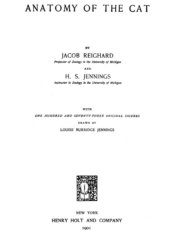
Fig. 26.—Frontal Bone, Medial Surface.
a, frontal spine; b, transverse ridge; c, surface applied to the ethmoid; d, vertical plate of medial border.
The ventral surface is concave and smooth over its caudal one-half and helps to form the cranial part of the brain-case. It presents slight ridges and depressions for convolutions of the cerebrum. At its narrowed middle region the ventral surface is marked by a thick transverse ridge (Fig. 26, b). Caudally the ridge descends by a gentle slope to the level of the ventral surface of the bone. The cranial end of the ridge is pierced by an oval foramen through which the frontal sinus (Fig. 43, m, m′), which lies within the ridge, communicates with the spaces in the ethmoid bone (nasal cavity). Craniad of the ridge the surface (Fig. 26, c) is rough and, together with the raised medial border of the bone and the orbital plate, encloses a rectangular space which in the natural state receives a portion of the labyrinth of the ethmoid. The ventral surface is marked at its medial edge by a thin longitudinal ridge which, when the bones are articulated, is continuous with one of the vertical lamellæ of the ethmoid.
The medial border forms a vertical plate (d), broadest craniad and roughened for articulation with its fellow of the opposite side except at its cranial end, where it articulates with the border of the nasal bone.
The caudal border is roughened, bevelled at the expense of the outer surface, and articulated with the parietal bone except at its ventral end, where it articulates with the alisphenoid.
The lateral border is smooth, and it is here that the orbital plate is joined to the frontal plate at right angles. Along its cranial two-thirds this union is marked by a sharp ridge, the supraorbital margin (Fig. 40, o) or arch. This ridge extends caudolaterad as a triangular projection, the zygomatic (or postorbital) process (Fig. 40, n), which is flattened on its cranioventral face near its extremity and forms part of the boundary of the orbital fossa. At its cranial end the lateral border articulates with the nasal and maxillary bones.
The orbital plate (Fig. 40, 5′) arises from the ventral surface of the lateral border of the frontal plate. It is directed ventrad, is smooth and concave on its outer surface, and forms the dorsal portion of the medial wall of the orbital fossa. Near its ventral border it bears the small ethmoidal foramen, for the artery of the same name.
On the caudal one-half of its inner surface (Fig. 26) it assists the caudal part of the dorsal plate in forming the brain-case. The cranial one-half of its inner surface is marked off from the remainder of the surface by a sharp irregular ridge which is for articulation with the cribriform plate of the ethmoid. Craniad of this the surface is marked by ridges and looks into the nasal cavity.
The cranial margin is produced dorsally in the form of a blunt triangular spine. Mediad of this spine the bone articulates with the lachrymal bone.
The ventral border articulates by its cranial one-third with the orbital plate of the palatine, and by its caudal two-thirds with the body and wing of the presphenoid.
Maxillary Bone. Maxilla
(Figs. 27 and 28).—The maxillary bone forms the cranial and lateral portions of the roof of the mouth. The bones of opposite sides meet craniad, but diverge caudad to enclose the palatal plates of the palatine bones. Each consists of a thick prismatic ventral portion or body (a) and a thin flat plate, the frontal process (b), extending dorsad from the cranial part of the bone.
Fig. 27.—Maxillary Bone, Lateral Surface.
Fig. 28.—Maxillary Bone, Medial Surface.
a, body; b, frontal process; c, infraorbital foramen; d, elevation for root of canine tooth; e, canine tooth; f, first premolar; g, second premolar; h, third premolar; i, molar tooth; j, zygomatic process; k, beginning of lachrymal canal; l, ridge to which the ventral nasal concha is attached; m, nasal crest of palatine process.
The body (a) has the form of a triangular prism whose broader dorsal face looks into the nasal cavity and orbit, while the ventral face looks into the mouth, and the lateral face toward the cheek. From the junction of the dorsal and lateral surfaces at the cranial end the large flat curved frontal process (b) passes dorsad, while the teeth are implanted along the border, alveolar border or process, formed by the junction of the ventral and lateral surfaces.
The lateral surface is continuous with the lateral surface of the frontal process and shows at the base of the frontal process on its caudal border the large infraorbital foramen (Fig. 27, c), for the vessels and nerves of the same name. Near the medial end of the surface is a cylindrical elevation (d) for the root of the canine tooth (e).
The ventral surface is smooth and looks into the roof of the mouth.
On the dorsal surface caudal and cranial halves may be distinguished. The caudal one-half enters into the floor of the orbit. The lateral edge of this portion is divided into two laminæ, between which is received the end of the malar bone. Caudad this edge is prolonged into the short dorsally directed zygomatic process (j). The cranial half of the dorsal surface looks into the nasal cavity and is separated from the caudal half by a sharp vertical lamina of bone which runs caudomediad from the base of the nasal process. To the dorsal edge of this lamina are articulated the lachrymal bone and a part of the palatine. At the point where the lamina joins the base of the nasal process a foramen is seen leading into a canal, the nasolachrymal canal (k). Craniad of the lamina the surface is concave. Where it becomes continuous with the inner edge of the frontal process there is attached to it a thin bone, the ventral nasal concha (or maxilloturbinal), which is rolled into an irregular spiral. The nasolachrymal canal opens ventrad of its cranial end.
The cranial third of this part of the bone projects further mediad than does the rest of the medial border, forming thus the broad palatine process. This is rough on its medial edge for articulation with the premaxillary and the palatine process of the opposite bone. This medial edge rises also dorsally into a low ridge, the nasal crest (m), which is roughened for articulation with the vomer. The caudal two-thirds of the medial edge articulates with the palatine bone.
The cranial end of the bone articulates with the premaxilla.
The caudal end is smooth.
The frontal process (b) presents on its inner surface, which looks into the nasal cavity, certain transverse ridges which are in relation with the ethmoid bone. Its outer surface is smooth. By its cranial border it articulates with the nasal bone dorsally and with the premaxillary bone ventrally.
Fig. 29.—Premaxillary Bone, Obliquely Craniolateral Aspect.
a, the three incisor teeth; b, palatal portion of the bone; c, nasal process.
Its dorsal end articulates medially with the nasal spine of the frontal bone, and caudally with the orbital plate of the same bone.
Premaxillary Bone. (Os incisivum BNA.) Premaxilla
(Fig. 29).—The premaxillary bones bear the incisor teeth and form the cranial portion of the roof of the mouth.
Each consists of an irregular, horizontal palatal portion (b) and of a perpendicular nasal process (c) which forms part of the lateral boundary of the nares and enters into the formation of the lateral wall of the nasal cavity.
The palatal portion has in its caudal border a deep notch for the foramen incisivum or anterior palatine canal, which lies between it and the maxillary and transmits blood-vessels and nerves. It articulates with the maxillary bone by this border.
The medial border is raised into a thin crest of bone which, besides forming the medial wall of the foramen incisivum or anterior palatine canal, articulates by its medial border with the bone of the opposite side, forming a sort of median trough (sulcus palatinus) which projects dorsad into the nasal cavity and receives the ventral border of the nasal septum. The caudal end of this border articulates laterad with the maxilla, dorsad with the vomer.
Its craniolateral border bears the incisor teeth (a).
The nasal process (c) presents three surfaces, all elongated and triangular; one, the medial surface, is smooth and concave and looks into the nasal cavity. Its dorsal border is rough for articulation with the nasal bone dorsad, and smooth ventrad where it aids in forming the nares.
The lateral surface is smooth.
The caudal surface is rough for articulation with the maxillary bone.
Nasal Bone. Os nasale
(Fig. 30).—The nasal bones fill the space between the nasal process of the premaxillary, the frontal process of the maxillary, and the nasal spine of the frontal bone (Fig. 39, 7). They thus form part of the dorsal wall of the nasal cavity near the middle line.
Fig. 30.—Nasal Bone, Dorsal View.
Each may be described as consisting of two elongated triangular lamellæ, one vertical, the other horizontal. The vertical lamella is curved slightly ventrad and has its apex directed craniad. It is applied by its medial surface against the vertical lamella of the opposite bone, the two thus forming a median vertical partition, the nasal crest (Fig. 43, 12), which extends ventrad into the nasal cavity and, by joining the dorsal edge of the lamina perpendicularis, helps to form the internasal septum.
The horizontal lamella is attached to the dorsal margin of the vertical lamella in such a way that its apex lies opposite the base of the vertical lamella. It helps to roof in the nasal cavity, and by its base forms a part of the dorsal boundary of the narial opening. By its lateral margin it articulates with the nasal spine of the frontal at its caudal end, with the frontal process of the maxillary at its middle, and with the nasal process of the premaxilla at its cranial end. The lateral angle of its base projects in a curved line which forms the dorsal part of the lateral boundary of the narial opening.
From the lateral border of the horizontal lamella a bony plate curves ventrad and mediad, enclosing a narrow fossa which receives a part of the ethmoid. This is the concha nasalis superior (nasoturbinal bone).
Ethmoid Bone. Os ethmoidale
(Figs. 31 and 32).—The ethmoid bone closes in the cranial cavity at its cranial end and extends forward into the nasal cavity, which it largely fills.
It consists of a median vertical portion, the lamina perpendicularis (Fig. 43, n; Fig. 42, p), forming a part of the nasal septum, of two lateral portions made of thin sheets of bone variously folded and united—the labyrinths (or ethmoturbinals), which fill the greater part of the nasal cavity; and of a transverse perforated plate, the cribriform plate (lamina cribrosa), attached to the caudal end of the lamina perpendicularis and the labyrinths.
The lamina perpendicularis (Fig. 43, n; Fig. 42, p) is a flat four-sided bone. By its caudal margin it is continuous with the cribriform plate; by its ventral margin it is enclosed by the halves of the vomer; by its dorsal margin it unites with the crest formed by the vertical portion of the nasal bone craniad and with the vertical lamina of the medial margin of the frontal caudad, while its cranial margin is continued into the septal cartilage of the nose. Its lateral faces are smooth and free.
The lamina cribrosa or cribriform plate (Fig. 42, o) is elongated heart-shaped, with the apex of the heart ventrad. Its caudal face is concave and looks into the cranial cavity. It presents three irregular longitudinal rows of holes, one median and two lateral, for the passage of the olfactory fibres from the cranial cavity into the nasal cavity. Its cranial face is continuous along the medial line with the lamina perpendicularis, and at the sides with the labyrinths.
The notch in the heart is directed dorsad and receives the vertical lamina of the medial border of the frontal bone. The apex of the heart articulates with the cranial end of the dorsal surface of the presphenoid. Its lateral margins are articulated with the ethmoidal ridges on the medial surface of the frontal bone.
a, vomer; b, vertical cells of the labyrinth of the ethmoid; c, horizontal cell of the same; d, part of the ethmoid that forms the lamina papyracea; e, edge of cribriform plate.
The labyrinths (Figs. 31 and 32) are attached to the cranial face of the lamina cribrosa, one on each side of the lamina perpendicularis. Each is made of thin bony plates irregularly folded so as to enclose spaces, the ethmoid cells. In each may be distinguished a cranial portion (b), in which the cells are nearly vertical, and a caudal portion (c), in which the cells are nearly horizontal.
The medial surfaces are separated by a space from the lamina perpendicularis. This space is broadest along the junction of the horizontal and vertical portions of the labyrinth. There are thus formed two passageways which correspond to the superior meati of human anatomy.
The lateral surfaces come into contact with the frontal process of the maxillary and the orbital plate of the frontal bone. On the lateral surface of each labyrinth there is a thin irregular lamina of bone lying in a dorsoventral longitudinal plane and closing in some of the ethmoid cells laterally (d). A small part of this lamina, situated near the caudoventral angle of the bone, appears in the orbital fossa on the external surface of the skull between the presphenoid, palatine, and frontal bones or between the lachrymal, palatine, and frontal bones. Sometimes in the entire skull two such pieces may be seen, one in each of these positions. This corresponds to the lamina papyracea of human anatomy.
The dorsocaudal angle of each bone is received into the space between the orbital plate of the frontal and the vertical lamina of the medial border of the frontal. Its ventrocaudal angle is received between the cranial extensions of the lateral walls of the presphenoid, while its ventral surface is overlaid caudally by the expanded portion of the vomer, to which it is attached at its caudolateral angles.
Vomer
(Figs. 31 and 32, a).—The vomer consists of two thin laminæ of bone which ensheath the ventral margin of the lamina perpendicularis (or the cartilaginous plate which continues ventrad from this margin) and unite ventrad of it; the two thus form a trough open dorsad.
Each becomes horizontal near its caudal end and at the same time expands. The expanded portion lies ventrad of the labyrinth of the ethmoid, closing in some of its cells: its lateral angles are united with the labyrinths.
At its caudal end the bone articulates with the body of the presphenoid, and each half of it is produced caudad near the middle line into a triangular spine which lies ventrad of the body of the presphenoid. The horizontal portion of the bone helps to separate the olfactory and respiratory passages of the nasal chamber, while its vertical portion contributes to the formation of the nasal septum.
The ventral margin formed by the junction of the two halves of the bone is smooth and free caudad, but at its cranial end is broad and rough for articulation with the palatal processes of the maxillæ.
Palatine Bone. Os palatinum
(Fig. 33).—The palate bone or palatine bone consists of two portions, a horizontal or palatal portion (a) and a perpendicular or nasal portion (b), uniting at an angle of about forty-five degrees.
Fig. 33.—Palatine Bone, Dorsal View.
a, horizontal portion; b, perpendicular portion; c, maxillary spine; d, posterior nasal spine; e, sphenopalatine foramen; f, caudal opening of posterior palatine canal.
The horizontal portions (a) of the two bones are received between the maxillary bones and form the caudal and medial part of the roof of the mouth. Each is irregularly quadrilateral in form, with the caudolateral angle produced caudad into a long process which is continuous with the perpendicular portion of the bone. The lateral margin of the horizontal portion articulates over its cranial half with the maxillary bone. At about its middle a short thick maxillary spine (c) projects caudolaterad. The remainder of the lateral margin is directly continuous with the perpendicular plate of the bone. The medial margin is rough for articulation with the corresponding margin of the opposite palatine; the caudal angle of this margin projects caudad as the short posterior nasal spine (d). The caudal margin forms a free edge which bounds the choanæ; it passes laterally into the perpendicular portion.
The ventral surface (Fig. 41, 8) looks into the mouth. Near the middle of its craniolateral margin are two or more small foramina (Fig. 41, q) which form the cranial termination of the posterior palatine canal. The dorsal surface is smooth and looks into the nasal cavity.
The perpendicular or nasal portion (Fig. 33, b) of the palatine is thin and irregularly quadrilateral in form. It is attached by its cranial two-thirds to the dorsal surface of the horizontal portion. The outer surface is concave and looks into the orbital fossa. The inner surface is convex and looks into the nasal cavity.
The perpendicular portion is marked by two foramina just craniad of the middle. The larger dorsal oval foramen is the sphenopalatine foramen (e). The smaller ventral foramen is the caudal opening of the posterior palatine canal (f). From this opening the canal passes craniomediad, lying in the substance of the palatine bone; it opens on the ventral surface of the horizontal portion at the small openings previously described (Fig. 41, q).
By its cranial margin it articulates with the lachrymal bone. By its dorsal margin it articulates craniad with the orbital plate of the frontal: with the lamina papyracea at its middle, and with the body of the presphenoid caudad. The caudal half of the dorsal margin is partially divided into two lamellæ with a rough surface between them: this rough surface lies against the ventral surface of the presphenoid. The caudal margin articulates with the pterygoid portion of the sphenoid.
Lachrymal Bone. Os lachrymale
(Fig. 34; Fig. 39, 10).—The lachrymal bone is a thin pentagonal scale of bone filling the interval between the horizontal plate of the palatine, the maxillary, and the orbital plate of the frontal. Its outer surface looks into the orbit, its inner surface into the nasal cavity.
Fig. 34.—Lachrymal Bone of Left Side, External Surface.
Fig. 35.—Malar Bone of Right Side, Lateral Surface.
Fig. 34.—a, notch forming the beginning of the lachrymal canal.
Fig. 35.—a, ridge for origin of the masseter muscle; b, frontal process; c, zygomatic process.
Near the middle of its cranial border it is notched obliquely by a foramen (a), the beginning of the nasolachrymal canal.
Malar Bone. Jugal Bone. Os zygomaticum
(Fig. 35).—The malar or zygomatic bone is a flat curved plate of bone which forms the lateral wall of the orbit and together with the zygomatic process of the temporal forms the zygomatic arch. Its outer surface is smooth and marked by a longitudinal ridge (a) for attachment of the masseter muscle.
At its caudal end the bone is continued into two processes: one, the frontal process or orbital process (b), is a triangular spine of bone directed caudomediad; when the bones are articulated it lies opposite the zygomatic process of the frontal to which it is joined by a ligament (orbital ligament). The other, zygomatic process (c) of the malar bone, extends ventrocaudad and articulates with a similar process from the temporal to form the zygomatic arch above mentioned.
Its inner surface is smooth and looks into the orbit, except that of the zygomatic process, which looks into the temporal fossa.
Its cranial border is roughened at the expense of both surfaces and articulates with the maxillary bone. Its other borders are smooth except the dorsal border of the zygomatic process, which is roughened for attachment to the zygomatic process of the temporal.
The Mandible. Mandibula
(Figs. 36 and 37).—The mandible (or inferior maxillary bone) is composed of two halves which come together at the cranial end and form the lower jaw. At its caudal end each half articulates with the temporal bone at the mandibular fossa, and at its cranial end it joins the opposite bone, the suture being known as the symphysis of the jaw (symphysis menti) (Fig. 37, a).
Each half consists of a horizontal portion, the body (b), bearing teeth on one of its borders (the alveolar border), and of a vertical portion, the ramus (c).
The body (b) has the form of a flattened cylinder and has two surfaces and two borders. The lateral surface (Fig. 36) is smooth and presents near its cranial end a foramen (or sometimes two), the mental foramen (d), forming the cranial termination of the mandibular canal. At its caudal end is a deep fossa continuing on to the ramus, the coronoid fossa, or masseteric fossa (e).
Fig. 36.—Mandible, Lateral Surface.
Fig. 37.—Mandible, Medial Surface.
a, symphysis; b, body; c, ramus; d, mental foramina; e, coronoid fossa; f, mandibular foramen; g, angular process; h, coronoid process; i, condyloid process; 1, 2, 3, the three incisor teeth; 4, the canine tooth; 5, 6, the premolars; 7, the molar tooth.
The medial surface (Fig. 37) is smooth and has near its caudal end a foramen, the mandibular foramen (f), which communicates with the mandibular canal leading lengthwise through the bone to the mental foramen. The cranial end is roughened for attachment to the bone of the opposite side.
The ventral border is smooth and rounded; it ends caudally in a blunt point, the angular process (g). The dorsal (alveolar) border is slightly curved and bears the sockets (alveoli) for the teeth. It is continuous with the cranial margin of the coronoid process.
The ramus is divided into two portions, the coronoid process (h) and the condyloid process (i). The coronoid process (h) extends dorsocaudad as a thin plate of bone with smooth surfaces and borders. Its outer surface is partly occupied by the coronoid fossa (e). The condyloid process (i) has the form of a semicylindrical transverse piece of bone attached to the caudal margin of the coronoid process. It articulates with the mandibular fossa of the temporal bone.
Hyoid Bone. Os hyoideum
(Fig. 38 and Fig. 104).—The hyoid bone forms the support for the tongue and gives origin to muscles passing to the tongue and larynx. It also supports the thyroid cartilage (Fig. 104, 1).
Fig. 38.—Hyoid Bone, Dorsal View.
a, body; b, c, d, e, cranial cornu; f, caudal cornu; b, ceratohyal; c, epihyal; d, stylohyal; e, tympanohyal; f, thyrohyal.
It consists of a transverse bony bar, the body (Fig. 38, a) and of two cornua or horns attached to each end of the body.
The cranial cornu (lesser cornu of human anatomy) is the longer (Fig. 38, b-e). Each arises from the cranial face of the body at its lateral end, curves laterad, and then caudodorsad. It consists of four bony pieces movably united by cartilage.
The terminal piece is the tympanohyal (e); it is imbedded in the tympanic bulla just ventrad of the stylomastoid foramen. It is not therefore seen attached to the cornu after the latter has been separated from the skull. The other pieces become successively shorter toward the body, and are called stylohyal (d), epihyal (c), and ceratohyal (b).
The caudal cornua (f) (greater cornua of human anatomy) arise from the ends of the body. Each consists of a single piece of bone, the thyrohyal (f), which passes caudolaterad; its free end is united to a process of the thyroid cartilage (Fig. 104, 1).
The Skull as a Whole.
—In the following description of the skull as a whole the mandible, hyoid, and ear-bones are not included.
The skull forms a bony box which contains the brain and is produced craniad into the facial portion which encloses the nasal cavity and forms the framework of the face.
In dorsal view (Fig. 39) the skull presents a smooth convex surface, broadest caudad, with the two zygomatic arches (g) curving out some distance laterally. The following bones are visible in dorsal view: the occipital (1), interparietal (2), parietals (3), temporals (4), frontals (5), malar or zygomatic bones (6), nasals (7), maxillaries (8), premaxillaries (9), and lachrymals (10).
Fig. 39.—Skull, Dorsal Surface.
1, occipital bone; 2, interparietal bone; 3, parietal bones; 4, temporal; 5, frontal; 6, malar; 7, nasal; 8, maxillary; 9, premaxillary; 10, lachrymal, a, lambdoidal ridge; b, external occipital tubercle; c, sagittal crest; d, parietal eminence; e, line which forms the dorsal boundary of the temporal fossa; f, zygomatic process of the frontal; g, zygomatic arch; h, frontal process of the malar; i, supraorbital arch; j, nares; k, foramen incisivum or anterior palatine foramen; l, sphenopalatine foramen; m, zygomatic process of the temporal; n, infraorbital foramen; o, opening of lachrymal duct.
The caudal boundary of the dorsal surface is marked by the prominent lambdoidal ridge (a) which passes from the middle cranioventrad along each side to the root of the zygomatic arch: it is borne by the occipital and temporal bones. From the middle of the lambdoidal ridge a second ridge, the sagittal crest (c), passes craniad in the middle line across the interparietal bone: it varies greatly in extent, reaching in a very old and muscular cat to the cranial border of the parietals, while in kittens it does not exist. The most prominent portions of the skull in this region, just craniad of the middle of the parietal bones, are known as the parietal tubercles or eminences (d). A faint curved line (e) runs from the cranial end of the sagittal crest craniolaterad to the base of the zygomatic process of the frontal: it marks the dorsal boundary of the origin of the temporal muscle, and may therefore be considered the dorsal boundary of the temporal fossa. This fossa extends from its dorsal boundary as far laterad and caudad as the lambdoidal ridge (a), and as far craniad as a line connecting the tip of the zygomatic process of the frontal (f) with the frontal process of the malar (h). The temporal muscle takes origin from its surface.
The middle portion of the dorsal surface is formed by the frontals (5). Each frontal presents laterally a prominent zygomatic process (f), extending ventrolaterad toward a corresponding (frontal) process (h) of the malar bone. These two processes mark the boundary between the orbital fossa (craniad) and the temporal fossa (caudad). Craniad of the zygomatic process of the frontal a sharp margin separates the dorsal surface of the skull from the wall of the orbital fossa: this is the supraorbital arch or margin (i).
The cranial portion of the dorsal surface is formed by the maxillary (8), nasal (7), and premaxillary bones (9). Just craniad of the nasals, bounded ventrad and craniad by the premaxillaries, appears the large opening of the nares (j), leading into the nasal cavity.
The zygomatic arch (g) is formed by the zygomatic process of the temporal (m) and the malar or zygomatic bone (6). Each presents near its middle a prominent dorsocaudally directed process, the frontal process (h) of the malar bone. The zygomatic arch forms the lateral boundary of the temporal and orbital fossæ, which are separated by a line connecting the frontal process of the malar (h) and the zygomatic process of the frontal (f).
A portion of the floor of the orbit and the opening of the lachrymal canal (o) may also be seen in dorsal view; they are described in connection with the lateral surface.
The caudal surface of the skull is formed largely by the occipital bone (Fig. 17), surrounding the foramen magnum (Fig. 17, d). At the sides of the foramen magnum are the two prominent curved occipital condyles (e) for articulation with the atlas. Craniolaterad of the condyles, separated from them by a deep notch, are the jugular processes (f) of the occipital, closely applied to the caudal ends of the tympanic bullæ.
Dorsad of the foramen magnum are faint indications of a median ridge running dorsad, the external occipital crest (Fig. 17, i); this rises at its junction with the lambdoidal ridge to form the prominent external occipital tubercle (Fig. 39, b). The dorsal and dorsolateral boundaries of the posterior surface are formed by the lambdoidal ridge (Fig. 17, h; Fig. 39, a).
The lateral surface of the skull (Fig. 40) is much more complicated than the dorsal and posterior surfaces. Caudally the occipital condyles (a) and external occipital crest (b) are visible; dorsocaudad the sagittal crest (c).
Extending from the caudal end of the sagittal crest the lambdoidal ridge (d) is seen passing ventrocraniad to the tympanic bulla, thence craniad to the root of the zygomatic arch. In the ventral part of the caudal region the tympanic bulla (e) is visible with the jugular process (f) of the occipital pressed close against its caudal end. Just craniad of the jugular process the mastoid process (g) of the temporal rests against the side of the bulla. Beneath the cranial edge of this process is the opening of the stylomastoid foramen (h) for the seventh nerve, while just ventrad of the foramen is the small pit (i) in the tympanic bulla for the reception of the tympanohyal bone. Craniad of the stylomastoid foramen is the large opening of the external auditory meatus (j), leading into the middle ear.
Immediately dorsocraniad of the external auditory meatus the zygomatic arch begins as the zygomatic process (k) of the temporal bone. On the cranial surface of the base of this process is the deep mandibular fossa (l) for the condyle of the mandible. This fossa is bounded caudally by the prominent postmandibular process (m).
Fig. 40.—Skull, Side View.
1, occipital bone; 2, interparietal; 3, parietal; 4, temporal; 5, 5′, frontal; 6, malar; 7, sphenoid; 8, palatine; 9, presphenoid; 10, maxillary; 11, nasal; 12, premaxillary; 13, incisor teeth; 14, canine; 15, 16, 17, premolars; 18, molar. a, occipital condyle; b, external occipital crest; c, sagittal crest; d, lambdoidal ridge; e, tympanic bulla; f, jugular process; g, mastoid process; h, stylo-mastoid foramen; i, pit for tympanohyal bone; j, external auditory meatus; k, zygomatic process of temporal bone; l, mandibular fossa; m, postmandibular process; n, zygomatic process of the frontal; o, supraorbital margin; p, external pterygoid fossa; q, sphenopalatine foramen; r, orbital fissure; s, internal pterygoid fossa; t, hamulus; u, foramen ovale; v, foramen rotundum; w, optic foramen; x, opening of lachrymal canal; y, infraorbital foramen.
All that portion of the lateral surface of the skull which lies craniodorsad of the lambdoidal ridge may be divided (excluding the zygomatic arch) into three main parts, the temporal fossa, the orbital fossa, and the face. The boundaries of the temporal fossa have been given. The orbital fossa is bounded externally by a prominent semicircular ridge formed chiefly by the zygomatic arch, the zygomatic process of the frontal (n), and the supraorbital arch (o) of the frontal, which may be traced to the cranial root of the zygomatic arch. The orbital fossa may be considered to end caudally and ventrally at the level of the optic foramen (w); ventrad of it are certain smaller fossæ. Immediately ventrad is the long external pterygoid fossa (p), from which arises part of the external pterygoid muscle. This fossa begins at the sphenopalatine foramen (q) and extends caudad to the orbital fissure (r); it is separated by a ridge from the orbital fossa. Caudoventrad of the external pterygoid fossa and separated from it by a sharp ridge is the small narrow internal pterygoid fossa (s), which extends ventrad without interruption on to the surface of the hamulus (t) and caudad to within two or three millimeters of the tympanic bulla. From it the internal pterygoid muscle takes origin. The hamulus (t) projects caudoventrad in this region, forming a prominent feature in a lateral view.
Four foramina leading into the cranial cavity are visible in a lateral view of the skull, craniad of the tympanic bulla. The one nearest the bulla is the foramen ovale (u) for the third division of the fifth nerve; next craniad of this is the foramen rotundum (v) for the second division of the fifth nerve. These two foramina pierce the alisphenoid: just craniad of them, between the alisphenoid and the orbitosphenoid, is the large orbital fissure (r) (foramen lacerum anterius), which transmits the third, fourth, and sixth cranial nerves and the first division of the fifth. Dorsocraniad of the orbital fissure is the optic foramen (w), for the optic nerve.
Ventrad of the cranial portion of the orbit is the large sphenopalatine foramen (q), for the nerves and arteries of the same name. Just craniad of this is the small caudal opening of the posterior palatine canal, which passes through the substance of the palatine bone and opens on its ventral surface near its cranial margin. Just dorsad of the cranial root of the zygomatic arch is the opening of the lachrymal canal (x), while the root of the arch is pierced by the large infraorbital foramen (y), which transmits the infraorbital nerves and artery from the orbit.
The teeth (13-18), implanted along the alveolar border of the maxillary and premaxillary, form a prominent feature in a lateral view: they are described in the account of the alimentary canal.
The ventral surface of the skull (Fig. 41) is very complex. It is separated by the orbits into a caudal and a cranial portion, united by a narrow median trough-like part. Laterad of this trough-like part are visible parts of the orbit and the zygomatic arches, which do not properly belong to the ventral surface and have already been described.