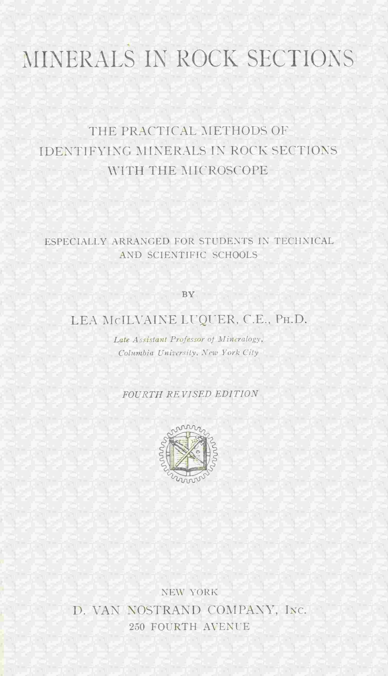
Fig. 52.—Augite, section parallel to ć axis showing prismatic cleavage, in leucite-basal. (From Cohen.)
Zonal structure (especially in augites) may be marked by differences in color or extinction, and in some basalts the crystals have the “hour-glass” structure.
Twinning.—Common, usually the twinning plane being the ortho pinacoid (100). Twin lamellæ may be noticed. Intergrowths occur with orthorhombic pyroxene and amphibole.
Color.—From almost colorless through green (diopsides, Na pyroxenes, etc.) to brown (augites); the red to brownish-red color of certain augites has been considered due to manganese. Yellow color very rare.
Index of Refraction.—n′ = 1.68 to 1.72 (α = 1.671 to 1.706, γ = 1.700 to 1.728), hence relief high and surface rough.
Cleavage.—More or less perfect parallel to prism of 87° 06′. Cleavage cracks distinct and numerous, but not generally running uninterruptedly through crystal, Figs. 12 and 52. Cleavage not so perfect as that of amphibole.
Parting.—Diallage and diopside have distinct parting parallel to ortho pinacoid (100), Fig. 53. Some crystals may show parting parallel to base (001).
Inclusions.—Tabular microscopic interpositions, similar to those in bronzite, may occur in diallage. The iron ores, apatite, etc., may occur in augite.
Polarized Light:
Fig. 53.—Diallage, cross-section.
Pleochroism.—Usually not noticed, and in general only appearing as different shades of the same color. In some cases (diallage, fassaite and Na rich augite) well marked, a and c green to yellowish green and b brownish to reddish-brown; hence pleochroism not intense in sections showing extinction angles. When Ti is present, violet parallel to b.
Crossed Nicols:
Double Refraction.—Strong (γ − α = 0.022 to 0.029), being stronger in the pale or colorless pyroxenes.
Interference Colors.—Second order, hence always bright tints.
Extinction.—Symmetrical in sections (through b axis) showing intersecting cleavage lines, in such cases bisecting the angles of the cleavage. In sections showing parallel cleavage lines, only parallel in ortho pinacoid (100) sections, in all other sections an extinction angle being observed. The maximum extinction angle is large, lies in the obtuse angle, varies with the chemical composition from 36° 30′ to 54°, and is only obtained when the section of the crystal is parallel to the clino pinacoid (010), Fig. 54, varying from this angle to 0°, when the section is parallel to the ortho pinacoid (100). In Ti and Na pyroxenes the inclined dispersion is so great that extinctions are not sharp, but instead a change takes place in the interference color from bluish to brownish.
Convergent Light: Axial plane parallel to clino pinacoid (010). Fig. 54. A cleavage flake parallel to ortho pinacoid (100) shows the emergence of an optic axis (orthorhombic pyroxene parallel to best pinacoidal cleavage would not show figure). Bxa.(c) Λ ć = 36° to 54° front. Axial angles large (2E = 70° to 112°). Optical character (+). The interference figures are distinct on account of the strong double refraction.
Alteration: May take place to chlorite, serpentine or amphibole (uralitization[92]), depending on the chemical composition and the conditions producing the change.
Fig. 54.—Diopside, clino pinacoid section.
Distinguished from:
(a) Orthorhombic Pyroxenes.—By extinction angle, the orthorhombic pyroxenes having always parallel or symmetrical extinction in sections parallel to a, b, or c, and by higher order interference colors. Also from hypersthene by absence of, or much fainter, pleochroism. Diallage and bronzite might be confused on account of pronounced pinacoidal parting, fibrous structure and inclusions; but may be distinguished by the presence or absence of extinction angles and also by the position of the optic axes relative to the best cleavage plates.
(b) Amphibole.—See under amphibole.
(c) Epidote and Chrysolite (Olivine). When light colored and granular, by examination in convergent light. The plane of the optic axes is parallel to the clino pinacoid (010), hence to the longitudinal axis and prismatic cleavage cracks, while in epidote it is at right angles to these directions and in chrysolite parallel to the base. Also yellow color is common in epidote but rare in pyroxene.
Remarks: Next to the feldspars pryoxene is the most common constituent of the igneous rocks. Diopside and fassaite (green) are found in contact rocks; also, what appear to be the same pyroxenes, in many eruptive rocks, as andesites, monzonites, etc. Malacolite (light green) is found in amphibolites and eclogites, where it may be associated with a greenish amphibole (smaragdite). Diallage (bladed and twinned) occurs in gabbros and pyroxenites. Common augite (brown) is found in the remaining basic eruptive rocks. In the schists the pyroxene is colorless.
Finally augite occurs as a secondary product resulting from the “magmatic resorption” of hornblende and biotite.
Chemical corrosion and mechanical deformation may occur. The green and brown augites when heated to redness on platinum foil may become red in color. In general the pyroxenes are not attacked by acids. H., 5 to 6. Sp. gr., 3.3 to 3.5. The sp. gr. of the pyroxenes is considerably higher than that of the amphiboles of similar composition, hence mechanical separations are possible.
Acmite (Ægirine) (Na pyroxenes).—Occur in green or brown, elongated prismatic crystals, often not very transparent and with marked pleochroism (like amphibole). Zonal coloring is common. When zonally intergrown with pyroxene the outer zone is ægirine. The elongation is ∥ a′ (distinction from amphibole whose elongation is ∥ c′). The index of refraction is higher than in the other pyroxenes (n′ = 1.792, α = 1.763, γ = 1.813) and the double refraction stronger (γ − α = 0.050). The extinction angle is small (5°) and the optical character (—).
The term Ægirine-augite may be used to describe a soda, pleochroic augite with a large extinction angle.
These pyroxenes are only found in the eruptive rocks rich in alkalies, as elæolite-syenite, phonolite, certain trachytes, etc.; hence are associated with elæolite, sodalite, leucite, etc. The small, second generation, crystals, in the ground mass of a rock, are always the richest in Na of the pyroxenes in that rock.
AMPHIBOLE, Hornblende, etc.
| Anisotropic. | Biaxial. | Monoclinic. |
| Elongation ∥ c′. |
Composition: RSiO3. R = Mg, Ca, Fe chiefly also may contain Al, Na, Mn. The Mg predominates over the Ca.
Fig. 55.—Hornblende, showing twinning between crossed nicols, in amphibole-biotite-granite. (From Cohen.)
Usual Appearance in Sections: Both in crystals and more or less irregular grains, often fibrous, Figs. 55 and 56, the habit varying with the chemical composition as follows:
Tremolite (Mg3Ca) and Actinolite ((MgFe)3Ca varieties), in long columnar to needle-like individuals, with no terminal planes or with frayed out ends. May be in dense aggregates.
Pargasite in well developed crystals.
Common green Hornblende (aluminous varieties) in crystals, compact grains or shreds.
Basaltic Hornblende (iron rich, aluminous varieties) in prismatic crystals of varying length, which may often show “magmatic resorption” (to augite and magnetite) around outer zone or throughout whole crystal.
Fig. 56.—Hornblende, section parallel to ć axis, showing prismatic cleavage, in hornblende-diorite. (From Cohen.)
Crystals are simple in form of prismatic habit, with prism angle 124° 30′. Cross-sections are acutely rhombic, generally with acute angles truncated, hence six-sided (pyroxene being eight-sided). Longitudinal sections are lath-shaped and fibrous structure may be noticed. Skeleton crystals may also occur, being very fine in certain pitchstones.
Zonal structure and parallel growth may be noticed in the amphiboles.
Twinning.—Frequent, parallel to ortho pinacoid (100). Twins dual, less often multiple, Fig. 55. Intergrowths with pyroxene and biotite occur.
Color.—From colorless (tremolite), through green (actinolite, pargasite and hornblende) to brown (basaltic hornblende). Yellow in some varieties and bluish in the soda varieties.
Index of Refraction.—n′ = 1.621 to 1.641 (α = 1.607 to 1.629, γ = 1.634 to 1.653) (1.719, in the basaltic hornblende), hence relief distinct and surface rough.
Cleavage.—Perfect, parallel to prism of 124° 30′. Generally appears in thin sections as sharp cracks crowded close together, Figs. 56 and 57. More perfect than in pyroxene.
Some of the long prisms (actinolite and tremolite) may show transverse parting.
Inclusions.—The iron ores, apatite, etc., may be found in hornblende.
Fig. 57.—Hornblende, cross-section.
Polarized Light:
Pleochroism.—All colored amphiboles show pleochroism, which in general is stronger the darker the color of the variety (actinolite and pargasite show but little). The absorption is very marked in the hornblendes, being greatest in the general direction of the cleavage lines in longitudinal sections. Marked differences in absorption are also characteristic of the mineral species biotite, tourmaline and allanite. Pleochroic halos (brownish) surrounding inclusions may be noticed.
Crossed Nicols:
Double Refraction.—Quite strong, but a little weaker than in pyroxene (γ-α = 0.019 to 0.027). Ferruginous basaltic hornblende has strong double refraction (γ-α = 0.072).
Fig. 58.—Actinolite, clino pinacoid section.
Interference Colors.—Second order, hence bright tints, but in darker colored varieties not so noticeable as in pyroxenes, due to the stronger absorption of parts of the light. The colors of basaltic hornblende are so high that they show no bright tints.
Extinction.—Always symmetrical in sections (through b axis) showing intersecting cleavage lines, in such cases bisecting the angles of the cleavage. In sections showing parallel cleavage lines, only parallel in ortho pinacoid (100) sections, in all other sections an extinction angle being observed. The maximum extinction angle lies in the acute angle and is much smaller than in pyroxene, varying with the chemical composition from 0°–20°. In hornblende, actinolite and tremolite 12°–20°, Fig. 58; in the basaltic hornblende 0°–10°. The maximum extinction angle is only obtained when the section of the crystal is parallel to the clino pinacoid (010), varying from this angle to 0°, when the section is parallel to the ortho pinacoid (100).
Convergent Light: Axial plane parallel to clino pinacoid (110), Fig. 58. Bxo·(c) Λ ć = 0°–20° behind. Axial angles large (2E = 77° to >180°). Optical character (−). Pargasite is (+).
Alteration: May take place to chlorite, talc, serpentine, asbestus, etc., depending on the chemical composition. Amphibole frays out and becomes fibrous during alteration, and may also lose color.
Distinguished from:
(a) Pyroxene.—By usually much stronger pleochroism in the colored varieties, and by cleavage and extinction angle. In pyroxene the cleavage (parallel to prism of 87° 06′) is less perfect; and the extinction angle is much larger, varying from 36° to 54°.
(b) Biotite.—By the extinction in the mica being always about parallel to the cleavage. Both have strong absorption, but biotite shows very slight pleochroism in sections parallel to the cleavage, and has only the one cleavage parallel to the base. Also the biotite has lower index of refraction and generally shows uniaxial interference figure.
Colorless tremolite may be distinguished from muscovite and talc by extinction angles, relief and lower order interference colors.
(c) Tourmaline.—By presence of cleavage, and by the fact that absorption is most marked about parallel to the elongation (also parallel to cleavage lines), while in tourmaline the absorption is strongest at right angles to the elongation.
(d) The Orthorhombic Pyroxenes.—By extinction angles, the latter having parallel extinction in all sections parallel to a, b and c, and by prismatic cleavage of 124° 30′. Pleochroism is strong in the colored varieties of both species, but in amphibole it appears more generally as a variation of the same color; while in hypersthene a change in color is often noticed, from brownish-red to greenish parallel to ć axis.
(e) Sillimanite and Cyanite.—See under the latter.
Remarks: Amphibole comes next to pyroxene in importance and distribution of the dark colored ferruginous rock-forming minerals. As a rule it occurs in rocks with a large percentage of SiO2, associated with quartz and orthoclase; while augite generally occurs in rocks of a basic nature, associated with plagioclase and little or no free SiO2. Furthermore amphibole contains hydroxyl and is therefore naturally found in the deep eruptive rocks; its place being taken by augite in the effusives. By application of heat hornblende changes to augite, while hydrochemical processes bring about the opposite result “uralitization.”
Tremolite and actinolite are found in contact rocks and crystalline schists, also as a result of the alteration of olivine into serpentine. Pargasite occurs in contact rocks. Common green hornblende is found in the plutonic rocks (Na poor and SiO2 rich), also in contact rocks and crystalline schists (amphibolites). Brown hornblende replaces the green variety in the basic plutonic rocks. Basaltic hornblende is found in many effusive rocks.
The hornblende crystals in eruptive rocks, being among the first formed constituents, have often suffered subsequent corrosion by the magma, giving rise to the dark border already mentioned. The brown primary hornblende in some rocks may be changed by a process analogous to “uralitization” into a green, reed-like hornblende. Mechanical deformations are found in massive and schistose rocks. Light green amphiboles, with weak pleochroism, may often be colored intensely reddish-brown and made strongly pleochroic by heating to redness on platinum foil. In general the amphiboles are not affected by acids. H., 5 to 6. Sp. gr., 2.9 to 3.3.
Glaucophane, Arfvedsonite, etc. (Na rich amphiboles).—Occur blue to bluish-green in color, with pleochroism and weaker double refraction than the other amphiboles. Extinction angles vary from 4°–6° (glaucophane) to 14° (arfvedsonite). They are found in contact rocks, crystalline schists, eclogite, etc.
For the rarer and less known members of the amphibole group, resource should be had to more elaborate works.
Uralite.—Pyroxene altered to amphibole, having the outward crystal form of pyroxene and the physical characters and cleavage of amphibole. The change usually commences on the surface and the uralite does not form a single compact crystal, but consists of numerous slender columns parallel to one another. These little columns or fibers have their c and [=b] axes parallel to the positions of these axes in the parent mineral. The color is green and the pleochroism weak to strong.
This change is called “uralitization” and results from hydrochemical processes. When the alteration is not complete, portions of the original pyroxene may be left, having all the characteristic optical properties of this latter mineral.
Anthophyllite, the orthorhombic amphibole, with always parallel extinction, is sometimes found in colorless to brownish, blade- to rod-like aggregates in crystalline schists and serpentine.
MICA GROUP.
| Anisotropic. | Biaxial. | Monoclinic. |
| May appear hexagonal or orthorhombic. | ||
| Composition: | Elongation (∥ cleavage) ∥ c′. | |
Biotite (black or ferro-magnesium mica) = (H.K)2(Mg.Fe)2Al2(SiO4)3, approx.
Phlogopite = a magnesium mica, near biotite, but containing little Fe.
Fig. 59.—Mica. A, biotite, showing hexagonal cross-section and zonal markings. Minette, Freiburg. B, Biotite, showing strong absorption parallel to cleavage and also zonal marking (P = plane of vibration of polarizer). Minette, Cumberland. C, Muscovite in bent shreds in gneiss.
Muscovite (white or potash mica) = H2(K.Na)Al3(SiO4)3, with some replacement by Mg or Fe.
Usual Appearance in Sections: Scales, which may be notched or jagged, with lateral sections lath-shaped; or shreds, Fig. 59 C. When distinctly crystallized (magnesium micas) the thin hexagonal plates have plane angles of 120°, Fig. 59 A. Phlogopite crystals may be extended in direction of ć axis.
Zonal structure not uncommon in the magnesium micas, Fig. 59 A, which may also have dark iron ore border like hornblende.
Twinning.—Common, generally parallel to base; seen in sections showing cleavage by variations in extinction, in basal sections by distorted interference figures.
Micas of different kinds often associated together in parallel position, also intergrown with hornblende, pyroxene, chlorite and quartz.
Color.—Depends on chemical composition. Biotites, brown, green or red to almost opaque. Phlogopites, colorless or yellowish. Muscovites colorless.
Index of Refraction.—n′ = 1.564 to 1.619 (α = 1.541 to 1.580, γ = 1.575 to 1.638), hence somewhat marked relief and surface varies in appearance from slightly rough to fairly rough. In polarized light the surface appears roughest when the cleavage cracks are parallel to the plane of the polarizer.
Biotite has more marked relief.
Cleavage.—Very perfect, parallel to base (001), Fig. 60. Basal sections show no cleavage, but all other sections show many sharp, parallel cleavage cracks.
Fig. 60.—Biotite, showing basal cleavage, in biotite-granite. (From Cohen).
For percussion and pressure figures, see reference given below.[93]
Inclusions.—May be arranged parallel to lines of pressure figure. Rutile needles, tourmaline, apatite, etc., common in magnesium mica. Zircon inclusions often surrounded by pleochroic halos.
Polarized Light:
Pleochroism.—Varies with the color, being very marked in the colored varieties (from pale yellow to chestnut-brown or black). The strong absorption, about parallel to the cleavage lines, is very characteristic of the colored mica, Fig. 59 B. Strong absorption is also noticed in hornblende, tourmaline and allanite. Absorption may even be noticed around inclusions (pleochroic halos) in colorless, non-pleochroic micas. Cleavage plates of biotite are not pleochroic unless the axial angle is large.
Crossed Nicols:
Double Refraction.—Very strong (γ − α = 0.034 to 0.058).
Interference Colors.—High order (third). May be very bright in thin sections of the colorless micas, and at times be so high in order as not to show any marked color tints. May not be noticeable in sections of the colored varieties, due to absorption of parts of the light.
Extinction.—About parallel to cleavage lines. Very small extinction angles may be noticed in biotites. Basal sections of biotite (the approximately hexagonal mica) usually appear isotropic.
Mottled appearance (like “Birds-eye” maple) characteristic, caused by distortion of the flexible laminæ during grinding. Most noticeable in sections inclined to cleavage and near position of extinction.
Convergent Light: Axial plane[94] and Bxa, practically at right angles to basal cleavage; therefore cleavage plates always show well defined interference figures, generally biaxial in character. The axial angles vary greatly, being usually small for biotite and phlogopite (may appear uniaxial) and large for muscovite (2E = 55° to 90°). Optical character for all micas (−).
Alteration: Biotites decompose quite easily, lose color and may become completely bleached, which appears to be due to a leaching out of the iron. May also alter to green chlorite, with a fraying out of the mica and a change to chloritic structure.
Phlogopites may alter to fibrous, scaly masses, apparently chiefly talc. “Sagenite” webs of rutile may accompany the alteration.
Muscovites are characterized by their freshness, and do not seem to suffer from weathering.
Distinguished from:
(a) Hornblende.—Magnesium mica has extinction about parallel to the cleavage, while hornblende may have extinction angles of from 0° to 20°. Both have strong absorption, but biotite shows very slight pleochroism in basal sections, which also give approximately uniaxial interference figures in convergent light.
(b) Tourmaline.—Magnesium mica shreds show absorption parallel to elongation, while in tourmaline the absorption is at right angles to elongation. There is also an absence of cleavage in tourmaline.
(c) Chlorite.—By strong double refraction, the very high order colors, however, being often not noticed. Chlorite also shows aggregate structure and is almost always greenish in color.
(d) Talc.—White mica by large axial angle of scales in convergent light and by micro-chemical tests. The distinction may be very difficult.
Remarks: Muscovite is a rare primary mineral in eruptive rocks, except in two-mica granite, etc. As a secondary mineral it occurs in dense scaly aggregates or as pseudomorphs after feldspar, nephelite, etc. It is frequent in crystalline schists and is probably also the mica in amphibolite and eclogite. Phlogopite is found chiefly in contact metamorphic limestone; and may be distinguished from muscovite by nearly uniaxial character and less sharp cleavage. Biotite is much more widely distributed, occurring especially in eruptive rocks, crystalline schists and contact rocks.
Chemical corrosion occurs in original biotite of porphyritic rocks, producing a “resorption border” of augite and magnetite. Mechanical deformations, producing bending and slipping along “gliding” planes (oblique to cleavage), are common to all varieties of mica and may produce change to chlorite. The muscovites, together with the feldspars, are the most characteristic minerals of dynamo metamorphic origin. Biotites and phlogopites are attacked by sulphuric acid at high temperatures. Muscovite is but slightly attacked by acids. H., 2 to 3. Sp. gr., 2.7 to 3.2. The specific gravity separation between the micas is difficult on account of the scaly nature of the minerals.
Other micas occur, some being alteration products of those already described. Among these may be mentioned:
Lithia Mica.—Both light and dark colored, occurring in granitic rocks and often only distinguished chemically from muscovite and biotite.
Damourite (Sericite) (hydrous K mica).—A secondary product usually in colorless, fine scaly aggregates in phillites, sericite-schists, etc.
CHLORITE GROUP.
Embracing the members of the Chlorite Group, commonly occurring in rocks.
| Anisotropic. | Biaxial. | Monoclinic. |
The minerals of this group usually appear uniaxial, and crystallize in part with hexagonal symmetry.
Composition: May be considered as isomorphous mixtures of H4(MgFe)3Si2O9 and H4(MgFe)2(AlFe)2SiO9 (Rosenbusch).
Usual Appearance in Sections: Minute, scaly aggregates, which may incline to radial grouping; or in minute grains as a pigment (veridite) in other minerals.
Twinning.—May be seen as in mica.
Color.—Generally green, varying from greenish white to dark green, rarely colorless or red.
Index of Refraction.—n′ = 1.567 to 1.589 (α = 1.560 to 1.585, γ = 1.571 to 1.596), hence no marked relief and only slightly rough surface.
Cleavage.—Like mica, very perfect; parallel to flat face, which is considered to be the basal plane. This cleavage may not be noticed, especially in secondary chlorite in rocks.
Polarized Light:
Pleochroism.—In green and yellow tints (green ∥ cleavage), being more marked in dark colored varieties. Basal sections are non-pleochroic, the mineral being practically uniaxial. Pleochroic halos may be seen.
Crossed Nicols:
Double Refraction.—Generally very weak (γ − α = 0.001 to 0.011).
Interference Colors.—Very low first order, gray or white, at times scarcely noticed. Anomalous colors, however, often seen (deep blue or brown.)
Extinction.—Plates parallel to cleavage generally appear isotropic or only show faint color. In other sections extinction is apparently parallel to the cleavage in the uniaxial type, but extinction angles may be noticed when type is biaxial. Complete extinction may not be noticed, due to aggregate structure.
Convergent Light: Plates parallel to cleavage show, at times, an indistinct interference cross, which may open into two hyperbolas, indicating biaxial nature of crystallization. Ax. pl. ∥ (010); Bxa. Λ ć = 0° to 15°; 2E variable. Optical character (±).
Distinguished from: Serpentine.—By general green color (serpentine, with exception of Fe rich variety, is colorless), pleochroism and frequent anomalous interference colors; but the distinction between these two minerals may be very difficult. Chlorite may resemble decomposed or green mica (mica has, however, strong double refraction). The different species in the chlorite group cannot usually be distinguished in rocks.
Remarks: The chlorites are essentially secondary minerals, derived from the aluminous silicates, biotite, augite, garnet, feldspar, etc. They are found abundantly in chlorite-schists, contact rocks, etc., and as pigment (veridite) in altered eruptive rocks. May occur as a primary constituent of eruptive rocks, often in parallel growth with biotite. Chlorides are acted on by hot hydrochloric acid, and decomposed easily by sulphuric acid. H., 2 to 3. Sp. gr., 2.6 to 2.96. A thin section heated to redness on platinum foil loses water and becomes opaque. Ferruginous varieties are turned reddish-brown to black by heating (serpentine, as it contains less iron, may give negative results with this test).
Delessite.—Found in sphærulites, filling cavities in amygdaloidal basic rocks, and in pseudomorphs. It appears to be much altered to other minerals.
TALC.
| Anisotropic. | Monoclinic (Pseudo-Hexagonal). | |
| Composition: H2Mg3(SiO3)4. | Elongation ∥ c′. |
Usual Appearance in Sections: In fine scaly, colorless aggregates. Sections, cutting across the scales, would show rod-like forms. Index of refraction only a little higher than balsam (n′ = 1.572, α = 1.539, γ = 1.589), hence no marked relief and only slightly rough surface. Cleavage perfect parallel to base, like mica.
Crossed Nicols: Double refraction very strong (γ − α = 0.050). Interference colors third order, like muscovite. Extinction parallel to basal cleavage lines. In convergent light, Ax. pl. ∥ (100), Bxa. ∥ ć; axial angle small (2E = small); optical character (−).
Distinguished from: Muscovite (with which it is easily confused) by micro-chemical tests, proving absence of alkalies and Al; and often by the more aggregate structure of the talc. Also by smaller axial angle of scales in convergent light.
Remarks: Found mainly in metamorphic schists, etc., always as a secondary product. It is insoluble in hydrochloric acid. H., 1 to 1.5. Sp. gr., 2.6 to 2.8.
EPIDOTE.
| Anisotropic. | Biaxial. | Monoclinic. |
| Elongation ∥ a′ or c′. |
Composition: Ca2Al2(AlOH)(SiO4)3, with some Fe replacing Al.
Usual Appearance in Sections: Columnar to thick tabular crystals, more or less elongated parallel to ortho axis [=b], Fig. 61, or in granular aggregates.
Epidote.
Fig. 61. Fig. 62.
Ortho pinacoid section. Clino pinacoid section.
Twinning.—May occur but rarely noticed. Irregular interpenetrations common with other members of the group; also parallel growths.
Color.—Colorless to yellowish (Fe poor) or yellow to greenish to yellow-brown (Fe rich).
Index of Refraction.—n′ = 1.751 but may be lower (α = 1.731, γ = 1.768), hence relief high and surface rough.
Cleavage.—Parallel to base (001), Figs. 61 and 62, imperfect parallel to ortho pinacoid (100). Basal cleavage cracks not very numerous and appear parallel to general direction of elongation.
Polarized light:
Pleochroism.—Varies with the color, being faint in the light colored varieties, but strong when the color is marked (Fe rich).
Crossed Nicols:
Double Refraction.—Variable, often very strong (γ − α = 0.037).
Interference Colors.—Variable, often high (third) orders. Intergrowths with other members of the group are clearly shown by the “flecked” interference colors.
Extinction.—Parallel to cleavage in sections parallel to [=b] axis. In other sections extinction angles vary, see Fig. 62.
Convergent Light: Axial plane ∥ (010), i. e., at right angles to the elongation of crystal and cleavage cracks, Fig. 61. Bxa.-(a) Λ ć= 3° behind. Basal cleavage flakes show the almost ⟂ emergence of an optic axis. Axial angles very large (2E > 180°). Optical character (−).
Alteration: Does not take place readily.
Distinguished from: Light colored Monoclinic Pyroxene.—By having plane of optic axes at right angles to cleavage cracks and direction of elongation; while in pyroxene plane of optic axes is parallel to parallel prismatic cleavage cracks or bisects the angle between intersecting cracks. Furthermore the yellow color is rare in pyroxene.
Remarks: Epidote is essentially a secondary mineral, resulting from the alteration of the feldspars and the ferro-magnesium silicates. It is found in crystalline schists (especially those containing hornblende), gneiss, gabbro, diorite, diabase, lime-silicate hornstones, contact rocks, etc. Epidote is partially decomposed by hydrochloric acid. The Fe rich epidote can be changed to an intense color by “glowing” in the air. H., 6 to 7. Sp. gr., 3.32 to 3.45.
Piedmontite (containing Mn).—Red in color. Very pleochroic, red to yellow. Found in crystalline schists, the porphyrite of Scotland, the famous “Porfido rosso antico” of Egypt and in certain Japanese mica schists.
ZOISITE.
Composition: Like epidote but without any Fe.
Usual Appearance in Sections: Similar to epidote or in columnar aggregates. Often intergrown with epidote.
Distinguished from epidote by general absence of color (colorless to yellowish) and pleochroism; by slightly lower refractive index (n′ = 1.699 to 1.720) and by much weaker double refraction (γ − α = 0.005 and less). The interference colors are very low order, gray to white, but anomalous colors are often seen (yellow or prussian blue). Extinction is in general parallel to pinacoidal cleavage cracks (except in clinozoisite).
The plane of the optic axes may be parallel or at right angles to cleavage cracks, and the optical character is (+).
Remarks: Generally a secondary mineral. Found in crystalline schists, amphibolites, contact rocks, eclogite, etc., and in “saussurite.” May be hard to distinguish from vesuvianite and apatite.
ALLANITE, Orthite.
Composition: Like epidote but containing cerium.
Usual Appearance in Sections: Similar to epidote in form; but distinguished by brown color (may be also almost colorless), strong pleochroism and absorption, and medium to weak double refraction (γ − α = 0.002 to 0.030). Lamellar twinning clearly seen on account of oblique extinction. When included in hornblende and mica it is surrounded by pleochroic halos. n′ = 1.78 about. Optical character (±). Usually perfectly fresh.
Remarks: Found as an accessory mineral in SiO2 rich eruptive rocks and connected crystalline schists, and (light colored) in amphibolite and eclogite.
TITANITE, Sphene.
| Anisotropic. | Biaxial. | Monoclinic. |
| Composition: CaTiSiO5. | Elongation ∥ a′.[96] |
Usual Appearance in Sections: Wedge-shaped crystals (in Na rich rocks, more prismatically developed); grains, which may be elongated; and aggregates of small rounded particles, which appear nearly opaque. Sections of crystals commonly acute rhombs, Fig. 63.
Twinning.—Occurs, the twinning boundary bisecting the acute angles of the rhomb (only noticed between crossed nicols), Fig. 64.
Color.—Reddish-brown to yellowish to colorless.