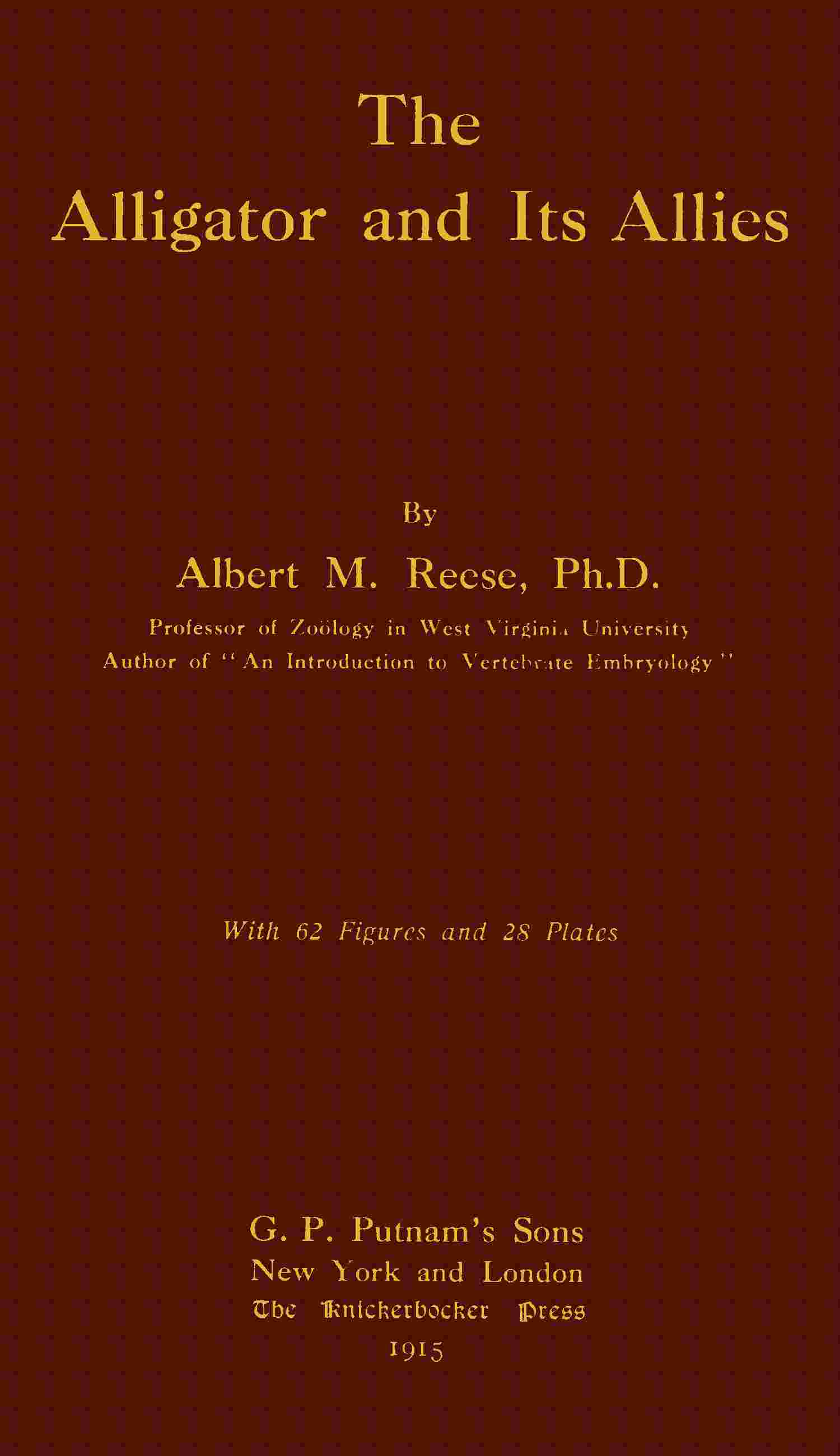
Fig. 25. Hyoids of an Alligator (Caiman latirostris) (TO THE LEFT) and of a Green Turtle (Chelone midas) (TO THE RIGHT). ×⁵⁄₈. (Brit. Mus.) (After Reynolds.)
The cartilaginous portions are dotted.
- 1. basilingual plate or body of the hyoid.
- 2. hyoid arch.
- 3. first branchial arch (anterior cornu).
- 4. second branchial arch (posterior cornu).
III. The Ribs and Sternum.
The Cervical Ribs.
As noted above, all of the cervical vertebræ possess ribs. The first rib, attached to the atlas, consists of a single, long blade projecting backward at an acute angle (Fig. 17, 8) as far as the middle of the fourth vertebra. As described above it articulates with the atlas at but one place. All of the other cervical ribs have two articular surfaces, a tuberculum and a capitulum, with a well-marked vertebrarterial canal between them. The ventral surface or capitulum articulates with a short process on the centrum; the dorsal surface or tuberculum (7) articulates with the transverse process. The third to seventh ribs are somewhat T-shaped, the stem of the T being the tubercle and head, while the cross arm of the T extends parallel to the axis of the neck (Fig. 17, 7). In the eighth rib the posterior arm of the T is elongated and projects out at a wide angle from the body; and in the ninth or last cervical rib this arm extends laterally as far as the vertebral portion of the thoracic ribs and has a cartilaginous tip.
The Thoracic Ribs
(Figs. 16 and 26). These are ten in number, the first eight pairs being connected with the sternum. The fourth may be taken as typical. It consists of a bony vertebral portion and partially ossified intermediate and sternal portions. The vertebral portion articulates with its corresponding transverse process by two surfaces, as described in connection with the thoracic vertebræ. In the first and second ribs only the tuberculum articulates with the transverse process, the head having a separate articular surface on the side of the centrum, as in the typical cervical rib. In the last thoracic rib the head and tubercle are not distinguishable from each other. Near the distal end of all the vertebral portions except the first and the last two ribs is a caudally projecting, partially ossified, uncinate process. The intermediate portion is present in all but the tenth rib, and wherever present, except in the ninth rib, it articulates distally with the sternal portion. The sternal portions extend medio-cephalad in a direction at right angles to the intermediate portion; the first two articulate with the sternum, the next six with the xiphisternal horns, and the ninth and tenth are missing.
Fig. 26. Sternum and Associated Membrane Bones of a Crocodile (C. palustris). ×¹⁄₃. (Brit. Mus.) (After Reynolds.)
The last pair of abdominal ribs which are united with the epipubes by a plate of cartilage have been omitted.
- 1. interclavicle.
- 2. sternum.
- 3. sternal rib.
- 4. abdominal splint rib.
- 5. xiphisternal horn.
The Abdominal Ribs
(Fig. 26, 4). While these ribs are membrane bones and are not homologous with the other ribs, they may as well be mentioned at this time. They consist of about seven V-shaped sets of slender bones, the point of each V being directed cephalad. Each V is made up of from two or five slender bones, the number and arrangement being subject to considerable variation. The last V of the series (not well shown in the figure) is considerably larger than the rest and is made up of four curved bones that extend around the anterior ends of the pubic bones and are united to them by a broad tough membrane. The first or most anterior V is united by a narrow membrane (not shown in the figure) with the membrane that extends between the xiphisternal horns. All of the V’s are more or less connected with each other by fibrous membranes. Since these ribs lie superficial to the recti muscles of the ventral body wall they are sometimes missing in carelessly prepared skeletons.
The Sternum
(Fig. 26). The sternum consists of the cartilaginous sternum proper (2), the xiphisternal horns (5), and the bony episternum or interclavicle (1). The latter is an elongated, flattened bone of somewhat spatulate outline, lying in the midventral line; it projects forwards to about the sixth cervical vertebra, while the anterior edge of the sternum is below the eighth cervical. Lying dorsal and lateral to the episternum is the flat, almost membranous sternum, to the posterior border of which the first two thoracic ribs are attached. The xiphisternum consists of two long, slender rods of cartilage; the anterior ends of these rods are in contact with each other and with the posterior border of the sternum; from this point they gradually diverge from each other as they extend caudad. A membrane extends between the horns as far back as the attachment of the last thoracic ribs.
IV. The Appendicular Skeleton.
The Pectoral Girdle and Anterior Limb.
The pectoral girdle (Fig. 27) is of a very simple type, consisting, unless the episternum (interclavicle) be counted, of but two bones, the scapula (s) and coracoid (c). The former consists of an upper, flat, paddle-shaped portion and a thicker lower portion which articulates anteriorly with the coracoid, and posteriorly forms about half of the notch-like glenoid cavity. The dorsal edge of the flattened portion is continued as a small, cartilaginous suprascapula. The coracoid is a flattened bone, wide at either end and narrow in the middle, so that in a dorsal view it is shaped like an hourglass. It is decidedly curved, with the convex side down. Its outer edge articulates with the scapula and is thickened to form the anterior border of the glenoid cavity. Its median end is attached to the sternum. Near its scapular articulation there is a well-marked foramen that passes entirely through the bone. The episternum (e) or interclavicle was described in connection with the sternum and ribs. There is no clavicle nor other coracoid elements.
Fig. 27. Pectoral Girdle and Anterior Limb.
c, coracoid; ce, centrale; cl, claw; e, episternum; h, humerus; m, metacarpals; p, pisiform; r, radius; r′, radiale; s, scapula; u, ulna; u′, ulnare.
The anterior limb consists of the usual parts,—the upper arm, forearm, and manus. The humerus (Fig. 27, h) is rather thick in proportion to its length; it has an elongated articular surface at its proximal end for articulation with the glenoid cavity, and a larger, somewhat bilobed surface for articulation with the radius and ulna. On its ventral side, near the proximal end, is a very prominent protuberance, the deltoid ridge. The ulna (u) is slightly heavier and longer than the radius and forms the greater part of the elbow joint and about half of the wrist joint. Its proximal end is considerably larger than the distal, but has no olecranon process. Its distal end articulates with the ulnare and pisiform. The ulna as a whole is slightly curved, while the radius is quite straight.
The radius (r) consists of a cylindrical shaft with enlargements of about equal size at the ends. The proximal end articulates with the side of the ulna and with the humerus; the distal end with the radiale.
The carpus consists of a proximal row of three distinct bones and a distal row of smaller and less fully ossified elements. Of the proximal row the radiale (r′) is much the largest bone. It is hourglass shaped, with the proximal end somewhat larger than the distal. Proximally it articulates mainly with the radius but also slightly with the ulna and ulnare. Distally it articulates with the centrale. The ulnare (u′), the second bone in size in the wrist, has about the same shape as the radiale but is much smaller. Proximally it articulates with the pisiform, radiale, and, apparently, with the ulna; distally it is in contact with the fused carpalia elements. The pisiform (p) is a small, irregular bone, articulating with the ulna and the ulnare; it is apparently connected by a long ligament with the fifth metacarpal but does not actually articulate with it. The centrale (ce) is a flattened, partially ossified element between the radiale and the first and second metacarpals. The distal carpal bones are represented by two irregular, partially ossified elements between the ulnare and the third, fourth, and fifth metacarpals.
The manus proper consists of five digits. The metacarpals (m) are of about the same shape, but vary in length and thickness; each consists of a cylindrical shaft with a slight enlargement at each end. The first digit or pollex has two phalanges, the second has three, the third has four, the fourth has four, and the fifth has three. The terminal phalanx of each of the first four digits is pointed, has a pair of lateral grooves, and is encased in a large, horny claw (cl).
The Pelvic Girdle and Posterior Limb.
The pelvic girdle is described differently by Wiedersheim and Reynolds; the bone called by the former the pubis, the latter calls the epipubis. The bone called by Wiedersheim the pubis takes no part in the formation of the acetabulum; the pubis of Reynolds helps form the acetabulum but is a very small, unossified structure. Gadow also calls the lower bone the epipubis. I shall follow Reynolds’s interpretation.
The ilium (Fig. 28, 1) is a heavy bone with a dorso-laterally projecting crest; medially it is firmly united to the sacral ribs (Fig. 18, 5) while its outer side forms the upper and greater part of the acetabulum. Its outer and lower border has two surfaces, the larger and more posterior articulating with the ischium, the other with the cartilaginous pubis.
Fig. 28. Pelvis and Sacrum of an Alligator (Caiman latirostris). ×¹⁄₂. (Brit. Mus.) (After Reynolds.)
- 1. ilium.
- 2. ischium.
- 3. true pubis.
- 4. epipubis (so-called pubis).
- 5. acetabular foramen.
- 6. neural spines of sacral vertebrae.
- 7. symphysis ischii.
- 8. process bearing prezygapophysis.
The ischium (2) is a slightly arched bone, its ventral end a flattened blade articulating with its fellow, its dorsal end enlarged and thickened to articulate with the ilium, pubis, and epipubis. This dorsal end, which forms the ventral side of the acetabulum, is divided into two distinct articular surfaces by a deep, rounded notch; the posterior and larger surface articulates with the ilium, the anterior surface about equally with the pubis and epipubis.
The pubis (3), which is much the smallest element of the pelvis, is a small mass of cartilage lying between the ilium above and the ischium below. It forms a small part of the anterior wall of the acetabulum.
The epipubis (4) is a slightly arched bone, somewhat enlarged at its proximal end where it unites with the ischium, and flattened out into a fan-shaped extremity, where it is united with its fellow and with the last pair of abdominal ribs by the broad, thin sheet of cartilage or fibrous tissue noted in connection with the abdominal ribs. As mentioned above, it is called by Wiedersheim and others the pubis. Near the center of the acetabulum there is a small foramen.
The posterior limb (Fig. 29) consists of the usual divisions—thigh, shin, and foot. The femur (f) is a bone of the same general outline as the humerus, though slightly longer and heavier. The head, for articulation with the acetabulum, is rather hemi-elliptical than hemispherical in shape, the long axis of the ellipse being vertical. The distal enlargement is of at least as great, if not greater, bulk than the proximal and shows some indication of a division into two articular surfaces. The ventral side of the femur near the proximal end shows a fairly distinct trochantal ridge.
The shin or crus is made up of two well-developed bones, the tibia (t) and fibula (fb), the former being somewhat longer and considerably thicker than the latter.
Fig. 29. Posterior Limb.
ca, calcaneum or fibulare; cl, claw; f, femur; fb, fibula; t, tibia; t³, t⁴⁻⁵, tarsalia; tb, tb′, tibiale-centrale; IV, V, 4th and 5th metatarsals.
The tibia consists of a cylindrical shaft with enlargements of about equal size at the ends. The proximal end forms most of the knee joint, the distal end articulates with a tarsal element said by Reynolds to represent the fused astragalus and centrale, by Wiedersheim called the astragalus, and said to represent the united tibiale, intermedium, and centrale (tb, tb′). The fibula articulates by a small enlargement at its proximal end with the femur, and by an enlargement of about equal size, at its distal end, with the fibulare or calcaneum (ca), and with a small facet on the above-mentioned tibiale-centrale element.
The tarsus is much modified and consists of four elements, in two rows; those of the proximal row are much larger than the two distal elements. Articulating with both tibia and fibula, as mentioned above, and with the first metatarsal and one of the distal tarsalia, is the large and irregular tibiale-centrale element of Reynolds (tb, tb′). In the tarsus here shown it consists of two elements. Post-axial in position is the calcaneum or fibulare (ca), articulating with the preceding tarsal element, with the fibula, with the rudimentary fifth metatarsal, and with the distal tarsal element said by Reynolds to represent the fourth and fifth tarsalia. The calcaneum is extended caudad into a prominent knob quite like the heel of the higher mammals.
The two distal tarsal bones are small; one is said by Reynolds to represent the first three tarsalia (t³), the other (t⁴⁻⁵) the fourth and fifth. Wiedersheim says one of these bones represents the first three tarsalia, the other the fourth. In the tarsus here shown these two elements are fused.
The foot has five digits, though the fifth is small and consists merely of a small, distally pointed metatarsal bone. According to Wiedersheim this fifth metatarsal is fused with the fifth tarsalia. The metatarsals of the first four digits are long and progressively more slender from the first to the fourth; each is distinctly enlarged at the ends. The first digit or hallux has two phalanges, the second has three, the third has four, and the fourth has four. According to Reynolds, the fourth toe has five phalanges; the figure here shown, which was drawn from nature, has only four on the fourth toe; the latter is the number given by Bronn for the crocodiles. The terminal phalanges of the first three digits are large and pointed, with the same lateral grooves noted in connection with the fore foot; each is sheathed in a horny claw. The four fully developed digits of the pes are nearly twice as long as the corresponding digits of the manus, but they are not proportionately thicker.