Fig. 209
Floating bodies—“joint mice”—from knee-joint. (Lexer.)
Symptoms.
—Rice-grain bodies may be suspected in cases of chronic tuberculosis and often in arthritis deformans, while in many instances they may be felt gliding beneath or between the joint structures. A perfectly loose floating body will produce symptoms which are quite distinctive. They consist of sudden and intense pain, with such muscle spasm as to fix the joint and prevent its use, thus “locking it.” Occurring at the knee the individual is instantly disabled, but usually learns by some peculiar manipulation, with or without assistance, to “unlock” the joint, and after a few moments to resume its use. Such a complaint as this should always suggest the condition. Patients who have had it for a long time learn how to avoid it as well as how to relieve it, and will often discover and be able to indicate to the surgeon the existence of a movable body, and even to describe its usual resting place.
Partial or complete dislocation of a semilunar cartilage in the knee is usually the result of traumatism, a distinct history of which can generally be obtained. It may not have been discovered at the time, owing to swelling or tenderness, but will produce its peculiar symptoms later, i. e., after use of the joint is resumed. Here, again, so long as it remain in proper position, it interferes but little; with a misstep or sudden movement, however, the patient is seized with sudden and painful disability. Here the movable cartilage may be felt projecting near its proper location. In such cases as these it is movable only to a certain extent and makes no free excursion about the joint. When not detected it may be suspected from the description which the patient gives of his seizures.
Diagnosis.
—So far as diagnosis is concerned, when a movable body can be felt all doubt is set at rest. When it cannot be discovered its existence may be inferred with an accuracy proportionate to the patient’s description of his difficulties.
Treatment.
—The treatment of rice-grain bodies is essentially that of the chronic hydrarthrosis and probably tuberculous condition which have led to their formation. It will consist usually in arthrotomy, with thorough irrigation; often in some form of arthrectomy. With the larger floating bodies, the “joint mice,” the most radical measures are the best. In most of these instances there will be some degree at least of hydrarthrosis. The joint cavity being distended and relaxed, the indication for arthrotomy is the more urgent, since it will permit also of irrigation or of dry sponging, with the same benefit with which analogous intraperitoneal conditions are treated by the same measures. The joint may be opened by a sufficiently ample incision, through which the foreign body or bodies may be removed. The operator should not be satisfied with mere removal of one, but should make a thorough search for others which may have escaped previous detection.
Perhaps no operative measure in surgery better illustrates the advantages of asepsis. This operation, which now can be done with impunity, was in the pre-antiseptic era one which had a discouraging fatality, death resulting from septic infection in about 40 per cent. of cases.
FOREIGN BODIES IN THE KNEE-JOINT.
“Joint mice” are of sufficient frequency and significance to justify brief separate consideration. According to Connell these may be grouped as follows:
- Those composed of foreign material, fatty tissue, fibrous tissue, etc.;
- Those composed of bone, cartilage, or of a mixture of the two.
Among the many explanations offered are the following:
- Dry arthritis, with overgrowth of the margins of the cartilages;
- Bony growths, separation from their attachments;
- Infarct of the articular cartilage, with final separation;
- Plate of bone formed outside of the joint and then invaginated;
- Calcification or chondrification of enlarged synovial fringes;
- Irritation and growth of embryonal cartilage or bone cells in the synovial fringes;
- Concretions whose nuclei are clots, torn fringes, or some foreign body;
- Some portion of the articular cartilages broken off by injury, or damage and subsequent separation.
Injury figures largely in the opinion of most of the authorities, it being well established that an injured portion of articular surface may become subsequently detached by a fatty necrosis, spoken of by König as osteochondritis dissecans, or by Paget as “quiet necrosis.” Others imagine that these floating bodies are rarely of traumatic origin.
Symptoms are usually marked and significant. There is sudden sharp and shooting pain, sometimes so severe as to cause faintness. Along with this there is “locking” i. e., fixation of the joint, usually in the flexed position, probably due to the entanglement of the floating body between the articular surfaces or between the bone and the capsule.
Fig. 210
Ankylosis of hip with contracture of knee, following post-scarlatinal arthritis.
It is the smaller rather than the larger bodies which give the most acute symptoms. This “locking” may last for only a few moments or for a number of hours and may or may not be followed by acute effusion. When with the above symptoms the presence in the joint of a movable mass can be made out diagnosis is complete. Some patients discover the movable body in their own joints before they go to the surgeon.
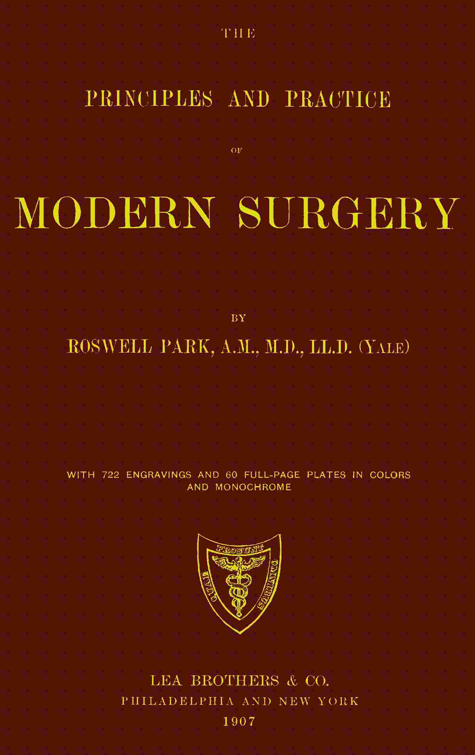
When the diagnosis is established the removal of the offending material is imperative. In the pre-antiseptic era this was an extremely hazardous operation. It is now one involving only theoretical risks. These bodies are sometimes extremely movable and slip about within the joint in a manner to almost defy removal even after the joint cavity is open. If such a body can be felt and fixed by digital pressure, or by the method of “stockading” suggested by Andrews some years ago, i. e., fixation by forcing sterilized pins into the tissues around it so that it cannot escape, it is then an easy matter to cut down upon it and remove it. Otherwise incision may require to be sufficiently ample to permit insertion of a finger and the general exploration of the joint before it is encountered. These bodies sometimes exist in small numbers, and it may be possible to remove several through a single opening. If the joint be opened and explored it should be done thoroughly in order that nothing may escape. After removal the capsule is closed with buried sutures, the balance of the wound closed as usual, and the limb then dressed upon a splint with absolute fixation for several days, in order to ensure physiological rest (Fig. 209).
ANKYLOSIS.
The term ankylosis implies angular deformity, but is used to designate partial or complete fixation of joints, such fixation being usually accompanied by more or less deformity or displacement. It is a name for a condition and not for a disease, but is always produced by the latter or by injury. The term itself implies nothing as to the nature, extent, or appearance of the exciting cause. The actual cause may have been disease of the joint, of the tissues around it, or may have been the result of injury rather than of infectious or other active disease.
For convenience we speak of fibrous, false, or pseudo-ankylosis, and of that which is bony or actual. A more accurate use of terms would lead us to refer to the former as contracture rather than true ankylosis.
Contractures are the result of acute, usually septic intra-articular and peri-articular processes, where muscle spasm is a pronounced factor and where the intensity of the process has more or less weakened the joint structures. The profession is hardly in the mood to accept acute rheumatism as an infectious process. If true or not the acute rheumatic affections are frequently followed by fibrous ankylosis with contractures. Disfigurements of this kind are often produced as the result of the surface lesions of severe burns or ulcerations, followed by cicatricial contraction and the formation of dense bands and scar tissue. This is a condition which can always be foreseen and which should be guarded against with very great care. (See Treatment of Burns.) Contractures also occur as the result of certain diseases of the spinal cord, either as the result of active contraction of one set of muscles, or of paralysis, by which the opposing muscles are deprived of resistance and thus draw the limb out of shape.
True ankylosis is sometimes fibrous, sometimes osseous, and occasionally both combined. The older the case the more probable is actual osseous union of joint surfaces. Bony ankylosis implies a sharply destructive type of arthritis, which may have been originally of pyogenic, gonorrheal, or tuberculous character, or else indicates a series of very slow ossific and calcific changes, such as are connected with the osteo-arthritis already described. Many of these cases are to be referred to lesions of the cord, and many of them are of polyarticular character. Fig. 195, illustrating one of the cases of so-called “ossified men” under the writer’s observation, will portray a series of lesions of this kind, most of the vertebral as well as the other joints being involved in an absolute osseous union.
Fig. 211
Bony ankylosis of hip. (Ransohoff.)
Fig. 212
Bony ankylosis of knee. (Ransohoff.)
Fig. 213
Bony ankylosis of hip with deformity. (Ransohoff.)
When a joint is stiff bony ankylosis may be inferred. So long as there is any motion possible it is essentially of the fibrous type. The condition is one easy of recognition, and is seen in all degrees of completeness. In many instances joint fixation is accompanied by adhesions of tendons and tendon sheaths, while as time passes all the structures around a joint thus fixed become less movable and more stiffened. Even the patella may become firmly attached to the bony surface upon which it normally rests, and thus interfere with motion of the knee almost as much as though the femur and the tibia were alone involved. Occasionally one of the acute exanthems is followed by contractures of a joint, with or without actual joint lesions, by which when neglected distressing deformities are produced; such, for instance, as partial flexion and fixation of the knees, or such stiffening of the hips as to prevent the thighs from being separated. While in such cases stiffening cannot always be prevented, deformity at least can be if suitable measures instituted sufficiently early.
Figs. 211 and 212, from Ransohoff, illustrate osseous union in the hip and the knee, while Fig. 213 illustrates the deformity which may be produced by contractures and ankylosis at the hip.
The following tabular presentation of the types of ankylosis will perhaps convey the greatest amount of information in small space:
| Ankylosis, true and false | - | Peri-articular | - | Capsular | ||||||||
| Extracapsular | - | Tendinous | ||||||||||
| Tendovaginal | ||||||||||||
| Muscular | ||||||||||||
| Articular | - | Synovial | ||||||||||
| Cartilaginous | ||||||||||||
| Osseous | ||||||||||||
Murphy has prepared the following table of the types of arthritis which lead to some of these varieties, and which may be classed as follows:
| Arthritis | - | (a) Primary hematogenous fibrous arthritis | ||||||||||||||||||
| (b) Dry fibrous arthritis. Non-traumatic | ||||||||||||||||||||
| (c) Traumatic fibrous arthritis | - | With fracture into joint | ||||||||||||||||||
| Without fracture (contusion) | ||||||||||||||||||||
| (d) Suppurative | - | Hematogenous | - | Cryptogenetic | ||||||||||||||||
| Metastatic | - | Typhoid | ||||||||||||||||||
| Scarlatina | ||||||||||||||||||||
| Pyemia | ||||||||||||||||||||
| Gonorrhea | ||||||||||||||||||||
| Traumatic | ||||||||||||||||||||
| Extension | - | Osteitis | - | Tuberculous | ||||||||||||||||
| Osteomyelitic (infective) | ||||||||||||||||||||
| Peri-arthritis (phlegmon) | ||||||||||||||||||||
| Panarthritis | ||||||||||||||||||||
| (e) Ossifying arthritis (primary) | ||||||||||||||||||||
| (f) Static adhesive | ||||||||||||||||||||
Treatment.
—The best method of treatment should be determined by the original character of the exciting cause, the duration of the condition, the amount of deformity present, and the degree of joint fixation. That which will be possible if done early will be useless if not resorted to until the case is old and chronic. In every acute or subacute condition which may threaten ankylosis every possible precaution should be taken to prevent it. If ankylosis be inevitable it should occur with the limb in the most suitable position. At the elbow, for example, this will be the right-angle position; at the knee, one with the leg almost completely extended. In the lower extremity traction with weight and pulley will serve a useful purpose in many instances, either to overcome a threatening condition or to improve one actually existant. Mechanical measures (i. e., use of various splints or forms of orthopedic apparatus) will sometimes be of great use. These may be arranged for the purpose of providing absolute rest, with fixation in a desirable position rather than in one which is undesirable, or they may be made with such devices as shall permit of frequent change of position.
The mildest operative measure which can be practised in these cases is manipulation, either gentle and frequent, combined with massage, or more violent and painful, such as requires anesthesia for its performance. The question of when to resort to these manipulations is one calling for the soundest judgment, as on one side the surgeon faces the possibility of setting up a renewed and more or less acute disturbance, and on the other of seeing a joint gradually stiffen, perhaps in a bad position. There is also a third difficulty, i. e., the necessity for continuing motion in order to prevent the re-formation of adhesions, and this in spite of the fact that it may be intensely painful to the patient. Fortunately, however, the use of nitrous oxide anesthesia usually permits this to be done as often as may be necessary with a minimum of discomfort.
Firm, fibrous ankylosis will be attacked with great hesitation by the experienced surgeon. Even though he may succeed in restoring the limb to a better position, he may feel quite positive that the patient cannot undergo the pain of the subsequent frequent handling. With bony ankyloses he may feel that nothing short of radical measures will suffice. Here it is rarely a question of restoring motility but rather of overcoming deformity. At the knee a wedge-shaped portion of the joint may be removed, its angle corresponding to the angle of deformity, and thus a crooked leg may be restored to the straight position; in fact, with a raised heel under such a limb it may be made almost as useful as ever. At the hip one may do a subcutaneous osteotomy, dividing the femoral neck either with chisel or with a small and protected saw, and then bringing the limb down into the normal position of extension, allowing the bone to repair itself, and effecting improvement only in position, or, by constantly moving it, securing a false joint; or a more formal exsection may be made and by removing the head of the femur and clearing out the acetabulum a degree of motion may be established at this point. At the wrist, elbow, and shoulder-joint resections will usually give good results if the operation be performed before the muscles have almost disappeared by atrophic processes.
Danger attaches to the performance of the so-called bloodless operations, in that there is a possibility of laceration of nerve trunks or of large vessels which may have become fixed in the condensed tissues and be torn with them. There is more danger of this perhaps at the knee than in other joints, and ruptures of the popliteal vessels and nerves have been repeatedly reported. The first attempt in breaking up such a joint should be to increase the degree of flexion. If by efforts in this direction the tissues can be first released, then there is less danger of their yielding when extension is made. Another danger which threatens in all resistant cases, and especially in elderly people, is fracture of bones. The writer has seen the upper end of the tibia as well as the neck of the humerus yield under these circumstances. In the latter event one should endeavor to prevent bony union, and thus to gain a false joint in place of the original.
In regard to the nature of the operative attacks upon the above types, the following is copied from Murphy:[33]
| A. Extracapsular disease | - | 1. Tendon elongation (tendoplasty). | |
| 2. Tendovaginitis (exsection of sheath). | |||
| 3. Cicatrices (removal). | |||
| B. Intracapsular | - | 1. Adhesive synovitis (exsection of capsule). | |
| 2. Replacement by aponeurosis or muscle. | |||
| C. Osseous | - | 1. Disconnect bones. | |
| 2. Remove neighboring bony processes or prominences. | |||
| 3. Liberate soft parts. | |||
| 4. Prevent subsequent bony contact. | |||
| 5. Interpose tissue to form hygroma or fibrous surface. | |||
| D. Joints suitable for operation. | - | 1. Mandibular. | |
| 2. Hip. | |||
| 3. Shoulder. | |||
| 4. Elbow. | |||
| 5. Knee. | |||
| E. Technique | - | 1. Flap formation (skin flap with fascia, or muscular). | |
| 2. Exposure of ankylosed area. | |||
| 3. Osseous separation. | |||
| 4. Transplantation and fixation of interposition flap. | |||
| 5. Replacement of bone. | |||
| 6. Fixation of parts. | |||
| 7. Drainage. | |||
| F. Subsequent treatment | - | 1. Passive motion | |
| 2. Active motion. | |||
| 3. Forced traction. | |||
[33] Journal American Medical Association, May 20, 1905, p. 1573.
To the various expedients which may be adopted for making stiffened joints more useful may be given, in a general way, the term arthroplasty. A variety of mechanical contrivances have been resorted to in the past, operators hoping to be able to secure, for instance, a movable knee instead of one which is stiff. Artificial joints, made of celluloid, ivory, etc., have been used for experimental purposes, but while occasionally they have given good results in animals, they have rarely been satisfactory in man. For the prevention of re-adhesion, plates of celluloid, thin metal, gutta-percha, rubber, etc., have been used. These are either wrapped around a bone end or are used for lining a bone cavity, and rapidly accumulating experience is showing that this may be done with great benefit.
Thoroughness of operative work is one of the important contributing agents to the securement of wide range of motion, especially in complete removal of synovial membrane, capsule, and ligaments. Soft parts should be liberated thoroughly. Of the materials which can be interposed between bone ends in order to prevent reunion, muscular aponeurosis, with a certain amount of fatty tissue, makes the best material for interposition. When aponeurosis cannot be secured, then muscle should be tried, with some fat, as the former flattens out and undergoes structural changes, with conversion into fibrous tissue.
It should be represented to the patient as a legitimate scientific experiment, and in such a way that no matter what may happen no blame can be attached to the operator. In general it may always be stated that the older the lesion the less satisfactory will be any measure of treatment except possibly resection and arthroplasty.
ARTHRODESIS.
This term applies to the intentional production of ankylosis in a joint previously healthy or nearly so, with the intention of stiffening a useless limb and thus enhancing its usefulness. The measure applies mainly to those cases of infantile paralysis, with loss of control of the knee or ankle, or both, when by stiffening the limb it can be made to serve the purpose of a crutch. It is the last resort in this direction when there is no possibility for tendon grafting. Long confinement of a limb in a fixed dressing will lead to considerable stiffening of the joint, yet a joint so immobilized lacks that firmness of support called for in cases above mentioned. Therefore when it is desired to perform arthrodesis the joint is usually opened and more or less of its articular surface removed, the intent being to produce the effect in the shortest time and in the best way. It can be better attained by a removal of articular surfaces with the saw and the apposition of fresh bone surfaces to each other, their retention being ensured either by sutures (tendon or wire) or accurate fixation in plaster of Paris. Under these circumstances drainage should not be necessary, and limbs can be completely enclosed in a fixed dressing.
MAJOR OPERATIONS ON JOINTS.
Aside from arthrotomy and partial or complete arthrectomy, as above mentioned, the latter, including removal of synovia or cartilage, and perhaps curetting of bone foci, the formal resections or excisions of joints remain to be considered. The latter is the preferable term, as it is meant to include removal of the component parts that enter into the construction of joints, while the term resection implies rather the removal merely of portions of bone.
Joint excisions are practised especially for the following purposes: (a) To atone for the result of old unreduced dislocations; (b) in certain compound dislocations, with or without fracture; (c) in certain comminuted fractures where there is no prospect of recovery with useful joints; (d) in the destructive forms of acute arthritis where the entire joint is disorganized and the bone ends carious; (e) in tuberculous arthritis or panarthritis, with or without suppurative complications; (f) in occasional instances of disabling osteo-arthritis; (g) for relief of ankylosis, either for improvement of position (knee) or restoration of motion; (h) occasionally after gunshot injuries. Excisions required by the exigencies of traumatisms should be promptly done. If the case be complicated with septic infection the prognosis is much less favorable. For convenience of description excisions may be classified as primary, intermediary, and secondary. According to the joint involved, as at the knee, the purpose underlying the operation is to effect an absolutely rigid bony ankylosis.
The development and perfection of the general method of joint excisions is a matter of but little more than a century. Previous to that time amputation was almost the only resort when destruction had occurred. The most prominent surgeons in the early development of the measure were Park, of Liverpool, and Moreau, of France. During the latter part of the past century Ollier, of Lyons, greatly improved the technique by demonstrating the importance of the periosteum and by introducing the so-called subperiosteal methods. This is of great value in uninfected cases. It is a mistake, however, to endeavor to save periosteum which has become involved in the tuberculous process; in fact, in the presence of tuberculous disease we cannot be too radical in the removal of all affected tissue.
In the so-called subperiosteal method the operator endeavors, so far as possible, to preserve the periosteum of the parts exposed to attack, and, if possible, the capsular ligament as well. Thus at the elbow the capsule, if not diseased or obliterated, should be preserved, the osseous tissue being shelled out from within, so far as possible. The less, then, the connections between the capsule and the periosteum are disturbed the better. The French apply to this method the term “subcapsular periosteal.” When the bone covering can be preserved new bone is easily formed to replace that which has been lost, especially during adolescence, while the preservation of the capsule, with its ligamentous connections, affords a better joint cavity than will the substitute which results from natural processes. Furthermore the surrounding tendons are less disturbed and the condition remains more like the original. Nevertheless one does not exsect healthy joints, and the method is not always easy nor even possible of performance. It will suffice to say that it should be adhered to only as far as circumstances may justify or permit.
Surgeons, however, have not been satisfied with the older methods, and have endeavored to still further enhance motility in operated joints. (See above—Arthroplasty.) To this end the interposition of muscle, fascia, or of foreign membrane has been suggested. Thus, after removal of the head of the femur a strip of fascia lata may be interposed between the raw-bone surface and the cavity of the acetabulum, being fastened there by catgut sutures. In the shoulder a similar procedure has been carried out, utilizing a strip of deltoid muscle. At the elbow a piece of the pronator radii teres may be detached and fixed by sutures to the brachialis anticus. In every case the method should be adapted to the demands made, the intent being to cover divided bone ends with tissue which will prevent osseous union, as it is known to do in many cases of fracture where such interposition produces non-union. In so far as one attempts here to imitate conditions which are considered undesirable in certain other traumatisms, Murphy has done more than any other American surgeon, both in the experimental and clinical study of this subject. (See above.)
For the joints below the hip and shoulder the bloodless method will facilitate operative work. In case of a septic joint, however, it would not be advisable to apply the elastic bandage below and then over and around the joint, as by the pressure thus made some septic material may be forced into the absorbents. In clean cases the rubber bandage is a great advantage to the operator. It has this objection, however, in that hemorrhage which does not occur during the operation has to be checked after its conclusion, and I have often thought it advisable to avoid the use of the bandage and to secure vessels as they are divided, in order that when bleeding has once ceased there be no fear of its recurrence later.
The question of drainage is one of importance. In a general way one may feel that in an absolutely clean case drainage is not required, save possibly a small opening for escape of blood. If practised at all it should be thoroughly done. Drainage tubes are often too small and do not permit the escape of either clotted blood or debris of injured tissue.
The after-treatment of excisions demands, first of all, physiological rest of the part involved, especially if, as at the knee, sutures or other expedients for maintaining apposition have been inserted. When motion is sought there will soon come a time when passive motion can be begun. This will vary with the size of the joint and the magnitude of the procedure. Actual rest should be maintained until firm wound healing has been secured. Passive motion is then begun, to be practised daily, the sensation of the patient being the guide as to the range of the movement and extent of manipulation. Thus, after exsection of an elbow with prompt union of the wound passive motion should be begun in about two weeks, but it should not be begun for a month if the joint has been thoroughly disorganized and the cavity is still discharging. Motion should be begun as early as is considered feasible in order to guard against a false joint.
The remote consequences of joint excisions are usually very satisfactory. The best results are obtained in the young, i. e., those whose tissues are still undergoing natural changes and whose bones are growing. In the course of time, by condensation of surrounding tissues, a new joint capsule is formed, its interior smoothed off, apparently covered with endothelium and filled with a sufficient amount of fluid, similar to that of normal joints, to serve the purpose; in this way a new joint becomes gradually substituted for the old, which serves the original purpose, in a surprising and gratifying way. Even in those of advanced years a satisfactory result is often obtained. It is often necessary to afford some support, by which too great a range of motion may be avoided; thus at the elbow the result at first is what may be called a “flail-joint,” which permits much undesirable lateral movement. This can be avoided by having light leather corsets fitted to the forearm and arm, connected by two lateral hinged braces. This being constantly worn, and no motion permitted which is not an imitation of the normal, the parts in time adapt themselves to the purpose, so that all apparatus can after a while be removed.
Excisions, like amputations, may be practised and the general methods learned on the cadaver, but their actual performance in the presence of extensive disease will be found to be a different procedure from that learned upon the dead body. For reasonably representative cases typical operations can be devised, with explicit directions. It is not advisable to try to do such work through too short incisions. A long incision heals as kindly as one shorter and affords more room for operative work. The incision should be so planned and executed as to afford the maximum of exposure with the minimum of damage to important structures. The region of the great vessels is avoided in all the classical operations, while nerve trunks, if exposed, are retracted and kept out of harm’s way. After the knife has once laid open the joint it is used but little except for the division of resisting structures, e. g., ligaments. The greater part of the work is then done with elevators, or periostomes with reasonably sharp edges and sufficiently broad surface, so that the periosteum can be divided with the latter and separated with the former to the necessary extent. Obviously epiphyseal junctions should be spared whenever possible, especially in the young. To remove an entire epiphysis is to materially impair the later growth of the limb. In some of the most serious cases it will be found already loosened and lying as a sequestrum in the joint cavity. In this case it may be easily lifted out of place. Tendons should never be divided unless absolutely necessary. Incisions in their neighborhood should be so planned as to be parallel with their direction and permit their displacement without division. The sharp spoon should be employed for curetting the interior of a joint capsule or cleaning out a bone focus (erasion). A capsule involved in tuberculous disease should be completely extirpated. Diseased bone ends should be sufficiently exposed to permit of the use of an ordinary saw or a chain or wire saw.[34] Considerable force will often be necessary in making bone ends accessible for this purpose. The chisel is rarely used except in cases of bony ankylosis, where it is not possible to force bone ends through the opening in order to attack them with the saw. As remarked above, clean cases may be closed without drainage. Visible vessels should be secured, and, while a certain amount of oozing may be expected, if the part be enclosed in suitable compressive dressings and elevated, it need not cause alarm. The gentle application of an elastic bandage for three or four hours may afford additional security. It should not, however, be allowed long to remain. The terminal portion of the limb will always afford an indication as to the condition of the circulation. Should it become cyanotic or cold the dressing should be renewed and the wound examined promptly.
[34] Wyeth’s “exsector” is an admirable substitute, especially at the shoulder and hip.
Special Incisions.
The Shoulder.
—A longitudinal incision suffices for most cases (Fig. 214). This may be made posteriorly between the fibers of the deltoid or anteriorly and externally over the bicipital groove. It is better to separate the deltoid fibers than to divide them, although they may be divided. Should the straight incision afford insufficient room another incision at right angles will afford ample access. The capsule, having been exposed, is opened, the wound widely separated with retractors, the arm rotated through a wide arc, while with a stout knife the capsular ligament and the various muscular attachments around the neck of the bone are divided. The greater and lesser tuberosities, with their muscles undivided, should be retained, when circumstances permit. The head of the bone, being freed, is dislocated and forced out through the wound, where it may be seized with large forceps and removed with a saw. The higher the bone is divided the better. Every other consideration, however, should be sacrificed to removal of all foci of disease. The capsule may then be extirpated and the glenoid cavity thoroughly cleaned out with a sharp spoon. Should the case be one of serious infection it is advisable to make a posterior opening, even through the deltoid, for purposes of thorough drainage. The greater part of the first incision is to be closed with sutures, the arm dressed in a comfortable position, with the elbow at a right angle, and the patient allowed to be up and around as soon as he feels in the mood for it.
Fig. 214
Excision of the shoulder: A, regular incision; B, supplementary. (Ollier.)
The Elbow.
—Here a variety of methods have been advised, and the extent of the operation must depend, to some degree at least, on the nature and extent of the condition which necessitates it. Partial excisions have been recommended, though in the writer’s experience incomplete operations often give less satisfaction than those which are complete. However, when it is a question of removing callus or displaced bone fragments, which, after fracture into the joint, impair its function, then partial resections may be serviceable.
Fig. 215
Fig. 216
Fig. 217
Excision of the elbow-joint: A, von Langenbeck; B, Ollier.
Excision of the elbow-joint: A, Nélaton; B, C, Hueter.
Osteoplastic method: A, by external incision; B, von Mosetig-Moorhof.
The essential incision is a long posterior one, which may be somewhat modified (Figs. 215, 216 and 217). It is essential here to avoid the ulnar nerve, which passes between the internal epicondyle and the olecranon, and the vessels and nerves in front of the joint. If it be made an inviolable rule to always keep close to the bone both of these dangers may be avoided. Ligamentous and muscular structures, among the latter the anconeus, should be spared as much as possible. After separating the joint surfaces thoroughly, by forced flexion, it is usually easier to force out the lower end of the humerus and first remove it, after which the upper ends of the radius and ulna are exposed and removed. When there is bony ankylosis it is preferable to divide the bones of the forearm first. The tendon of the triceps is not only detached from the olecranon, but divided by the first long incision. After concluding the incision, the capsule, if it remains, is to be closed with chromic catgut sutures and the end of the triceps tendon or some of its periosteal attachment united to the periosteum of the upper end of the ulna.
The arm is now fixed in the right-angle position and held comfortably to the body by a suitable sling.
The Wrist.
—It is rare that in disease of the wrist-joint this is found to be limited to a single bone of the carpus. Should an x-ray examination indicate such limitation then the focus can be exposed and cleaned by an incision upon the dorsum of the wrist, where it may seem best adapted for the purpose. Suppurative and tuberculous affections of the wrist usually necessitate removal of the carpal bones, including, possibly, the lower extremities of the ulna and radius. When the wrist-joint is involved it may be sufficient to remove the latter with the first row of the carpus.
Fig. 218 illustrates the incisions to be recommended for wrist resection, of which the Langenbeck line is to be preferred. Occasionally two lateral incisions, with through drainage, will better serve the purpose. It may be necessary to divide the short radial extensor, but this may be united again with suture. In most instances it is possible to retract the tendons to either side and thus clear the carpal region. By hyperextension the extensor tendons are relaxed and more room is thus made. The incision marked “A” combined with that marked “B” in Fig. 218, affords the best exposure when disease is extensive. The incision along the inner border of the wrist is made 5 Cm. above the styloid process of the ulna, and between the latter and the ulnar flexor down to the middle of the last metacarpal bone. Here the tendon of the latter muscle should be divided at its insertion and lifted out of its groove in the ulna. The collection of extensor tendons is then separated from the back of the wrist and lifted up, it being usually necessary to divide the unciform process of the unciform bone with forceps. The knife should be kept from the palmar surfaces of the metacarpal bones in order to avoid injury to the deep arch. After dividing the anterior radiocarpal ligament the carpus is extirpated through the ulnar incision. The ends of the ulna and radius are now easily accessible for removal with forceps or a metacarpal saw. The same is also true of the proximal ends of the metacarpals. After spreading the hand and forearm upon a flat splint drainage can be made to the desired extent and the wound closed.
Fig. 218
Excision of the wrist: A, Lister’s radial incision; B, Lister’s ulnar incision; C, Ollier; D, von Langenbeck.
Fig. 219
Excision of the hip: A, Sayre; B, Ollier.
So far as the hand and fingers are concerned little resecting need be done, the surgeon usually confining himself to the removal of sequestra or curetting of carious bone. In cases of compound comminuted fracture bone fragments may be removed; only in cases of lost or destroyed phalanges will amputation be necessary.
The Hip.
—In its structure the hip-joint is one of the simplest in the body. Although it lies deeply it is easily made accessible. Fig. 219 illustrates the incisions by which the joint is attacked for the purpose of exsection. If necessary either extremity of the incision can be extended or enlarged by a cross-cut. When the joint is disintegrated by disease, especially when partially dislocated, the parts will lend themselves to an easy and simple operation. When, however, the operation is done for ankylosis or for disease, by which great thickening and fixation have been produced, the measure may become difficult. For ordinary purposes the simplest method is to drive a sharp-pointed, strong-bladed knife directly down upon the neck of the bone from a point midway between the great trochanter and the crest of the ilium; then keeping the knife-blade in contact with the bone the incision is carried downward over the trochanter and along the shaft to a length making it sufficient for easy exposure of the bone and of the joint. Nothing is gained in these cases by trying to work through a short incision. A long one heals as readily and makes the operation more simple. It is as easy to make the entire incision in one cut as to divide the muscles layer by layer. The capsule of the neck of the femur being exposed by a wide retraction of wound margins, it is necessary next to divide muscular attachments to the great trochanter by raising the periosteum to which they are attached and saving both. To expose these insertions the femur should be rotated inward and outward, while the capsule is at the same time divided. The ligamentum teres, which offers a theoretical obstacle, usually disappears in the presence of any active disease and is scarcely ever encountered; it can be divided with curved scissors. Now by more or less powerful effort, including flexion and adduction to the extreme limit, with more or less rotation, the head of the bone is forced out from its socket and through the wound. Whether the bone should be decapitated with chain saw, metacarpal saw, or by the exsector of Wyeth will depend partly upon the freedom with which it can be exposed and on the equipment of the operator. It may be advisable to divide the neck with a chisel. The trochanter major should be preserved whenever its removal is not made imperative by the progress of the disease. The head and neck of the bone having been removed, the acetabulum is now more or less easily exposed, especially with retractors, and it should be cleaned with a sharp spoon. The capsule also should be removed, at least when the operation is done for tuberculous or other infectious condition. It is advisable to irrigate, then to wipe dry all the original joint surfaces and raw bone, and finally to cauterize either with pure carbolic or with zinc chloride, which should be washed away with the irrigating stream, the intent being to close the mouths of all the absorbents and prevent absorption from fresh exposure. Sinuses if present should be thoroughly excised, scraped, and treated in the same way. A drainage tube is usually preferable to the use of gauze.
The above is the method usually relied upon for hip exsection. Other methods have been devised, especially by anterior incision; of these the best probably is that of Barker. The cut is made along the outer border of the anterior surface of the sartorius and rectus, and through it the femoral neck is reached. By wide retraction the anterior surface of the joint can be completely exposed and opened, and through this opening the neck of the femur can be divided with a chain saw or chisel, before removal of the head from the acetabulum. The disadvantage of anterior incision is that pertaining to drainage. Nevertheless this can be obviated with capillary drains. Its advantages are that splinting and protection can be more perfectly effected, with less necessity for frequent interference. In other words it makes the subsequent care of the patient easier. Many English surgeons are in favor of it. Ollier devised a so-called osteoplastic excision, made through a curved incision with a downward convexity, the top of the great trochanter being exposed and divided with a chisel sufficiently to permit of its being turned up with the flap, and then being reunited to the main part of the bone after the removal of the neck and head. This method has its advantages in a limited number of cases, but it has not become popular in this country. It would seem to be an advantage to preserve the trochanter, although some surgeons remove it. So long, however, as disease is confined to the head and neck of the bone it is unnecessary to remove this projection.
The after-care of a hip excision is not an easy matter. Most surgeons prefer to maintain the limb in position by the aid of traction, with sufficient weight to overcome all muscle spasm. If the case be such that dressings need only be made at long intervals, then it matters little, but in a septic case in which there is considerable discharge the problem is sometimes a serious one. Various beds or suspension splints have been devised, consisting essentially of frames with cross-strips of stout material, upon which the patient lies. After raising the frame one or two of these strips are released and the parts exposed. This arrangement also permits of the easy management of a bed-pan. In young children a wire splint with a fenestrum, or a plaster-of-Paris spica or breeches with large opening cut opposite the wound, will often be serviceable. The tendency is rather toward adduction, and this should be overcome. Something will depend upon whether the surgeon is working for ankylosis or for a movable joint. In the former case a rigid dressing should be employed as soon as the condition of the wound permits. In the latter passive movement should be begun as soon as the wound is healed.
While the operation is usually performed quickly, and is not regarded as serious, it nevertheless has a considerable mortality, especially in the young and the aged, because of the conditions which necessitate it. After a complete exsection, even by the most ideal method and in the most ideal case, the limb remains somewhat shortened. This may be compensated by raising the heel of the shoe worn on the affected side. In severe cases it may be necessary to supply even two or three inches of artificial support for this purpose. Unless this is done compensatory spinal curvature will ensue.