CHAPTER LIV.
THE KIDNEYS.
CONGENITAL ANOMALIES AND DEFECTS OF THE KIDNEYS.
Recent embryological studies have established the fact, in regard to the kidneys, and given rise to the inference in regard to the other viscera, that the primary cause of congenital variations has much to do with the earliest development of the bloodvessels. The general inclination has been to view the vessels as following the organs. This should be reversed, as we are now learning that organs develop around the bloodvessels, and that so-called congenital variations arise from departures of vascular arrangement from the ordinary types. Without pursuing this subject further it is sufficient to say that, aside from defects of such character that the newborn infant can live but for a few hours or days, those which have most surgical interest mainly comprise variations in number and in size, including every possible combination, from absence of an entire kidney to horseshoe forms, and various anomalies of the ureters including defects and redundancies, double ureters, and the like. While supernumerary renal tissue or kidneys are extremely rare, the presence of supernumerary adrenal tissue in one or both kidneys (even in adjoining organs) is not uncommon. Here it may lead to the development of a distinct form of tumor, hypernephroma, which will be discussed later. The complication of absence of an entire kidney is sufficient to give it actual surgical importance, since it has repeatedly happened that the remaining kidney has been removed for disease, the inference being that its work could be carried on by its fellow, which proved to be lacking. This accident might be prevented by a careful cystoscopic examination. Nevertheless the rarity of this condition permits it to be almost excluded from ordinary consideration. After removal of one kidney the other undergoes compensatory physiological enlargement and does double duty, if indeed this has not already occurred.
Acquired defects may be due to intrinsic or extrinsic causes, e. g., disease within the renal structures or ureters, or lesions in adjoining organs and tissues, producing mechanical or other disturbances. Thus the functionating capacity of one or even both kidneys may be seriously compromised by either internal or external conditions, and it behooves the surgeon to estimate the degree of renal disability or inadequacy before operating upon either of these organs. On the other hand if the disease be confined to one kidney he may feel that it is doing so little good and so much harm that the patient will be really relieved by its removal. Nearly everything, then, depends upon a determination of the precise existing conditions. They should be ascertained by means of the catheter, the cystoscope, the microscope, and by the careful chemical study of the urine. These methods have been developed into a specialty of considerable complexity, but of great practical importance. The surgeon should not fail to employ them. If he is not familiar with the technique he should seek special assistance.
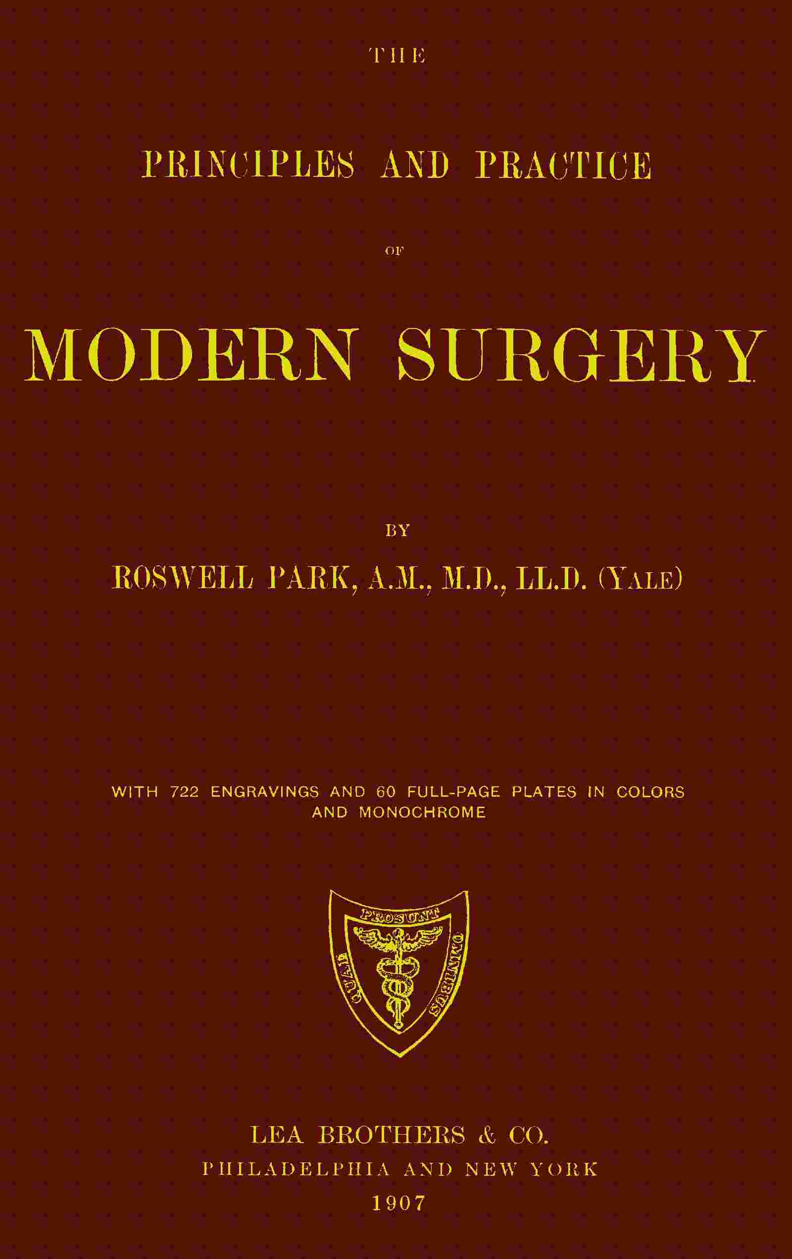
Fig. 632
Laceration fragmentation of kidney. (Güterbock.)
INJURIES TO THE KIDNEYS.
Although the kidneys lie in a protected position they are not infrequently injured, both by contusions and by penetrating wounds. From the latter blood will escape externally. In the former it can only extravasate when the cortex and capsule are torn, or escape through the ureter into the bladder, when it will be seen in the urine, which, however, may have to be drawn by the catheter on account of retention. Blood in the urine after a local injury denotes serious mischief inside the kidney or along the urinary tract. If continuing for several hours, but especially if accompanied by local indications, swelling or other evidences of extravasation, by muscle rigidity or by severe pain, with general symptoms, it should be assumed that these fluids are escaping into the perirenal tissues, perhaps into the peritoneal cavity, and that an immediate exploratory operation should be urged. When once this indication is clearly recognized the condition brooks no delay. The same is true of penetrating wounds. On general principles, with a patient in such a condition and showing no improvement, or especially if the reverse, exploration offers the safer course in by far the greater number of cases. The surgeon need only convince himself that such blood as the urine contains does not come from the lower tract, but rather from the kidney or ureter. Exploratory nephrotomy is by itself so harmless that one need never hesitate to urge it. A kidney found slightly lacerated may be repaired with sutures, while one found seriously disorganized should either be sutured and drained or totally removed, as the case may require. There is little room for doubt that it is better to institute such a measure early rather than to permit the dangers and even ravages of infiltration of blood and urine. In fact it may almost be laid down as a precept that every patient who has received an injury in the loin or flank and who repeatedly passes blood in the urine should be explored.
PAIN IN THE KIDNEY; NEPHRALGIA.
This is a vague term, implying pain or neuralgia in the kidney, and can refer only to symptoms, not to any particular disease. Yet it must be confessed that for certain cases of so-called nephralgia no physical cause is easily discovered. Pain in the kidneys—or, as patients will often say, in the back—may be associated with excess of oxalic and uric acids and salts in the urine, and is then relieved by a steady course of alkaline diuretic treatment, with plenty of fluid, the severe pain being combated with aspirin. Nephralgia may be expected in connection with many renal disorders, but the term should ordinarily be confined to cases of pain without known cause.
When such pain is uncontrollable and intolerable the indication is to make an exploratory operation, by which the kidney should be at least exposed, perhaps delivered upon the external surface of the body, and carefully examined. Its capsule should be split (capsulotomy), as Harrison and others have suggested, and if on palpation or needling (using a needle as a probe) there be any good reason for opening it this may be done, so that with the finger its pelvic cavity may be carefully explored, in order to find any previously unrecognized calculus or other surgical lesion. The mere operation of capsulotomy or capsule splitting has proved of such great value that I always practise this measure upon any kidney which for any reason it may seem wise to expose.
INFLAMMATIONS AND INFECTIONS OF THE KIDNEYS.
Under this head it is intended to consider (1) acute or subacute specific infections of the upper urinary passages, due to bacteria, with the effects of which we are familiar, i. e., septic, gonorrheal, and tuberculous lesions, and (2) chronic nephrites of irregular or uncertain type, for which operative treatment has been recently proposed.
Septic Nephritis; Pyelitis; Pyelonephritis; Surgical Kidney.
—Septic infection of the kidney is usually the result of a process ascending from the lower urinary passages, particularly when these are obstructed by calculus, tumor, prostatic enlargement, or ureteral stricture. It may follow catheterism either once or prolonged, especially when done without strict precaution; or the infection may come from the other direction via the blood stream, as in typhoid and various other fevers, the exanthems, and diphtheria. Gonorrhea is a frequent cause, acting insidiously and by a creeping invasion, with the intervention of a rather more abrupt cystitis. Nevertheless when gonorrhea is followed by pyemia and metastatic abscess these form early in both kidneys, and disaster quickly follows. These types of infection spreading upward along the ureters do not spare the pelvis of the kidney, but expend their first violence there. Beyond this they may extend to the renal tissue proper, where they set up a true nephritis, which may prove fatal.
Symptoms.
—Clinical symptoms do not vary greatly except in detail. They include fever, chills, and similar expressions of toxemia, with more or less pain in the kidney, down the ureter, and even referred to the ultimate distribution of the nerves sympathetically or anatomically involved, e. g., to the testicle on the same side, often with retraction of the scrotum, and down the thigh. There is a tendency to thamuria (frequency of urination) when the bladder is involved, as it always is sooner or later. Pus and mucus are recognizable in the urine by the naked eye, while a microscopic study of this fluid will reveal, from the character of the cells, the extent and type of the invasion. The tuberculous type will be considered separately. Suffice it to say that in this form, however pure may have been its original type, it becomes sooner or later converted into a mixed septic infection, with which renal abscess is often connected. The gonorrheal type is nowise clinically distinct, so far as the kidneys are concerned, but is to be recognized either by the microscope or by other clinical evidence.
Treatment.
—Such cases as the above may even perplex the surgeon, since they complicate many other surgical conditions. Yet if they go no farther than above described they are to be treated rather by internal methods, i. e., diluents, with hot-air baths, and especially by urotropin, the remedy of greatest value, while such drugs as aspirin, benzosol, sodium benzoate and the like, in moderate doses and at rather short intervals, may be administered to great advantage.
Renal Abscess; Surgical Kidney.
—The conditions above described do not necessarily nor often terminate with resolution. Not infrequently suppuration follows, with resultant abscesses, which may be solitary and possibly large, but are more likely to appear in multiple and perhaps punctate form. Should this condition occur in one kidney alone, it determines probably its ultimate destruction; if in both kidneys, the prognosis is very grave, since later, if not immediately, such a case will succumb to renal failure, due to the extra load put upon the portion still capable of secreting. It is to kidneys thus crippled by acute or subacute infections, with punctate abscess and similar lesions, that in the past the term “surgical kidney” was applied, because such kidneys were seen oftener in surgical than in so-called medical cases.
Brewer has recently called attention to a type of acute hematogenous renal infection, to which he has given an identity of its own. The possibility of renal infection through the blood has been long recognized, but it has been generally supposed to produce bilateral lesions. Of late, however, it has been shown that these may be unilateral, on account of the diminished resistance of one kidney as the result of previous disease or injury, among the former being calculus, renal retention, and floating kidney. While the colon bacilli are most frequently at fault the infection is often of the pyogenic or mixed type. It seems to be more frequent in women than in men. The symptoms are those of an acute infection, often ushered in by a chill, with sudden rise of temperature, sometimes followed by such marked remission as possibly to suggest malaria. The pulse ranges high. Abdominal pain is an almost constant symptom, although it is usually vague and often shifting or referred. Sometimes it will cause such a complaint as to lead to mistaken diagnosis in favor of an acute appendicitis. Occasionally it radiates along the course of the ureter. Tenderness in the costovertebral angle is nearly always present. Muscle rigidity is frequent but inconstant. There is nearly always a leukocytosis, with a percentage of about eighty polynuclears. Frequency of urination may accompany these cases, but they will ordinarily be diagnosticated by physical and urinary examination. The urine will usually contain albumin, perhaps with pus, and occasionally a few red blood cells. Urine obtained from the affected kidney by ureteral catheterization will contain more of these evidences of abscess than that from the other side. Brewer has had far better success in entirely removing the affected kidney than in exposing and simply draining it. He has thus done a great service in demonstrating the possibility of unilateral acute and suppurative disease of the kidney, where diagnosis is most obscure and the clinical picture one of acute general abscess rather than of local affection, showing as well that the more acute cases tend rapidly to terminate fatally unless promptly arrested by complete removal of the affected organ.
As we consider the above infections, with others yet to be mentioned, it becomes more necessary to appreciate those constituents and characteristics of the urine which have for the surgeon the greatest significance, and those methods of investigation which furnish him the promptest and most satisfactory results.
The following include methods in present use for determining renal capacity and function, i. e., the matters of greatest importance:
- 1. Catheterization of the ureters;
- 2. Cryoscopy of the blood and the urine;
- 3. Phloridzin test;
- 4. Chromocystoscopy;
- 5. The toxin test;
- 6. The test for electroconductivity (Kakells).
1. By cystoscopy, with ureteral catheterization, we determine whether urine is secreted by both kidneys or but one, while the secretion of each kidney may be separately collected and studied. Even this method leaves much to be desired. Though one kidney be actively diseased it may still contain sufficient tissue to make it partly competent for its purpose, and undesirable to remove; or an organ with very defective structure may, nevertheless, yield a certain amount of nearly normal urine. These, then, are aids to determine the character of the morbid process, and the information they furnish is valuable, but not always sufficient.
2. Cryoscopy, based upon the physiochemical law that the freezing point of the solution is proportionate to the number of molecules it contains, i. e., to its molecular concentration, has revealed that the blood of a person with severe kidney lesion freezes at a lower temperature, while the freezing point of his urine would be much higher than in a normal individual, because those materials which should have been excreted in the urine are, on account of impaired renal function, retained in the blood and do not get into the urine. The freezing point of normal urine varies from -0.09° to -2.3° C.; the freezing point of normal blood from -0.55° to -0.57° C. The reasoning employed in the method is sound, but the method itself difficult, requiring special apparatus and experience. Moreover, the limits of the possibility of error are such that this method alone should never be relied on. It is essentially a test of the ability of the kidneys to act as filters, but does not test their serviceability as secretory organs.
3. The phloridzin test is one of the most trustworthy for estimating the secretory function of the kidneys, as it shows how much active working epithelium remains in the organ. It consists in the subcutaneous injection of 0.005 Cc. sterilized phloridzin with an equal quantity of sodium benzoate, to hold the former in solution. The bladder must be emptied just before the injection is given. About an hour after its administration sugar should appear in the urine, if the kidneys are acting normally. If they are to be studied separately, catheterization of the ureters is necessary. The test is, of course, worthless in diabetic subjects. It depends upon the amount of sugar excreted, the time of its appearance, and the duration of its elimination. If no sugar be present the kidneys are seriously affected; if it be delayed, renal insufficiency is present. The average quantity of sugar eliminated during the first half-hour, when the kidneys are normal, is about 0.5 per cent. If the kidneys be diseased, this quantity is reduced by a half, and there is very little more secreted in the first than during the second half-hour. This valuable method is unfortunately difficult of application and requires minutely careful chemical tests.
4. Chromocystoscopy, introduced by Voelcker and Joseph, is perhaps the simplest of all methods of estimating renal capacity. 20 Cc. of a 0.4 per cent. solution of indigo-carmine is injected into the gluteal muscles. In fifteen or twenty minutes, if the kidneys be normal, the cystoscope will reveal dark-blue urine flowing from the ureteral orifices toward the median line, with a peculiar jet at regular intervals of about twenty-five seconds, and lasting for perhaps five seconds. There is both rhythm and force about this ejaculation. If the color be pale, the jet weak, or the rhythm irregular, the intervals prolonged or late, or if no flow whatever occur, there must be hindrance in the secreting and filtering structure of the kidney, or occlusion of the ureter. The results given by indigo-carmine in these cases are superior to those furnished by methylene blue, since it is not so much a solution as a mixture which is formed and ejaculated as such. Moreover, in passing through the body the indigo-carmine undergoes no reduction. By this method there is no necessity for catheterization of the ureter. One needs only to use the cystoscope with reasonable dexterity, and there is no necessity for chemical tests of separate specimens. The method is generally useful in cases where ureteral catheterization is made impossible by growths. It affords an easy means of differentiation, for instance, between ovarian cyst and hydronephrosis.
5. The toxin test is one only to be carried out by the use of animals, since it depends upon the amount of filtered urine required to kill an animal after injection into its veins, the number of cubic centimeters necessary to kill, divided by the weight of the animal, being called the urotoxic co-efficient. It has greater laboratory than clinical interest.
6. Electroconductivity of urine is of value in determining the capacity of the kidney for eliminating inorganic cells. It depends on the resistance offered by the urine to the electric current. It is complicated in method, requires special apparatus, and its results are still of questionable value.
For ordinary purposes the most trustworthy data for the surgeon who is not provided with ample laboratory facilities are afforded by an estimate of the amount passed in twenty-four hours, its specific gravity, its color and acidity, and by the presence or absence of albumin. The test-tube and the microscope then still afford satisfactory means of deciding those matters which the surgeon needs to know. If applied to urine collected separately from each kidney, they may be regarded as trustworthy. If catheterization be impossible, then it is advisable to inspect the ureteral orifices while elimination of indigo-carmine is taking place.
Hematuria.
—The significance of blood in the urine is rather that of a symptom than of a disease, although it should be admitted that there are occasional patients who lose blood in this way, more or less frequently, even periodically, without seeming to suffer in the least. Hematuria may also be present as an expression of vicarious menstruation. Again, blood may thus appear in scurvy and similar conditions, especially in tropical climates; in certain of the domestic animals its presence may be due to infection of the kidneys by macroscopic parasites (the so-called “black-water fever” of men and horses). Such cases as these are outside the pale of surgery. Nevertheless general experience has shown that many cases of hematuria, without perceptible changes in the kidney, have been benefited or cured by exploratory nephrotomy. Among the causes ascribed for these so-called “essential hematurias” have been incipient tuberculosis, renal retention from prostatic enlargement, congestion from venous obstruction (due to tight lacing or displacement from any cause), and even the congestion of chronic nephritis.
Treatment.
—When known or recognizable causes are absent, and the ordinary therapeutic agents, the special styptics (cotarnin), and such measures as hypodermoclysis with a 2 per cent. gelatin solution (see Control of Hemorrhage) have failed, an exploration may be advised. It is of the greatest advantage to be certain that but one kidney is involved, or it may be necessary later to operate on the second kidney.
Operative Treatment of Chronic Nephritis.
—The various changes included under this head are usually bilateral. The term implies a non-pyogenic infection of the renal bloodvessels, interstitial tissues, and glomeruli or tubules, which produce changes, often spoken of in this country as constituting interstitial parenchymatous or diffuse forms of nephritis, and inducing gross changes which cause the kidney to be spoken of as contracted, large, white, waxy, etc. Discussion of the pathology of these conditions here is out of place. They have all been grouped, most loosely, in common parlance as forms of “Bright’s disease.” Apart from the significance of albuminuria and the many terms implying peculiar features, the apparent hopelessness of many of these conditions, and the disappointment following internal treatment, finally led surgeons to attempt to ascertain what they could accomplish. It was in 1886 that Péan operated on a case of chronic nephritis with nephralgia and removed the kidney. Ten years later Harrison made three nephrotomies, and, though under a wrong diagnosis in each case, it was noticed that the symptoms all cleared up and that albumin disappeared from the urine. About the same time Newman showed that albumin and casts have often appeared in movable kidney, because of torsion of the vessels, and that they disappeared after nephropexy. Then Pousson, in 1899, reported some twenty-five cases of hematuria and nephralgic nephritis, operated upon by nephrotomy and nephropexy, with great benefit. In 1899, Israel was, perhaps, the first to formulate rules for nephrotomy for these conditions. In 1899, also, Ferguson claimed that chronic nephritis should be treated as are inflammations elsewhere, by relief of tension and even drainage. Meantime, Edebohls had been doing partial decapsulation and fixation in cases of so-called unilateral nephritis (the possibility of which is disputed by the best authorities, like Kümmel), and later extended his method to complete decapsulation (capsulectomy), with replacement of the kidney in its fatty bed, claiming that by and through the new adhesions thus produced new and more complete as well as additional blood supply was furnished, and that regeneration of the slightly altered parenchymatous tissue, as well as absorption of exudates, was produced. (Guiteras.) The fact that it seems now well established that these forms of chronic nephritis are always bilateral does not of itself affect the cogency of Edebohls’ reasoning, if it be otherwise correct.
Accurate diagnosis has much to do with this problem. Israel has shown that chronic nephritis is even more difficult of recognition in the living than in the dead, not only after ordinary examination of the capsule, but also after opening into the kidney. Age is not a serious contra-indication, and enlargement of the heart is said frequently to subside after these operations. If cardiac compensation be good operation is permissible, if not otherwise contra-indicated. Edebohls’ method is to anchor the kidney to the muscles of the back, whether it was previously movable or not. Primary healing is desirable, since “nephritics” do not bear suppuration well.
Indications for Operation.
—At present a satisfactory summary is impossible. It is of the first importance that operation should be undertaken early, since to wait until anasarca or other grave conditions supervene is to invite disappointment as the result of a procedure which is by many considered capital. The coincidence of pronounced disease of any other type would be a contra-indication. Bacteriuria, pyuria, etc., would perhaps make it more desirable rather than otherwise. Cases of operative toxemia (postscarlatinal, typhoid) and of cirrhotic type, without other contra-indications, are the most favorable. When a careful examination of the patient and the urine leads the surgeon to think that preparatory treatment may be of advantage, he should find therein almost his only excuse for delay, if operation is to be done. Low hemoglobin percentage should also lead to postponement.
Operation may consist of nephrolysis, or breaking down of adhesions, by which pain is frequently relieved, of decapsulation, of nephrotomy, and, finally, of nephrectomy, in case serious lesions are disclosed. It is doubtful if benefit is due so much to formation of new vessels as to a freer circulation of blood within the kidneys, with their consequent improved opportunity for repair and elimination. Guiteras, for instance, does not believe in total decapsulation, but in partial exposure of a sufficient area on the posterior kidney surface to assist in its fixation, if movable. Otherwise he considers that simple division of the capsule over the convexity will be sufficient. In cases of unilateral nephralgia and hematuria he advises nephrotomy, not so much as an approved therapeutic measure as for exposure, perhaps for revealing the possible existence of deep lesions.
Fig. 633
Acute pyelonephritis with multiple miliary abscess formation. (Israel.)
The recent reports from various surgeons concerning the value of renal decapsulation alone are by no means unanimously favorable, although a majority of writers are in favor of exposure of the kidney, capsulotomy and fixation, either by suture or tampon. Still, it does not seem at present justifiable to maintain that decapsulation can be expected to cure diffuse or deep-seated arteriosclerosis or degenerative processes within the kidneys.
The question of the suitable anesthetic is here one of importance. For reasons set forth earlier in this work, ether should always be avoided. If the operation be one that can be speedily performed, nitrous oxide gas alone may suffice. Otherwise it should be done under chloroform, preceded perhaps with ethyl chloride, or under somnoform.
Pyonephrosis.
—As a condition this is to be distinguished from ordinary abscess of the kidney, in that it implies the retention in the renal cavity or pelvis of pus with eventual destruction of kidney tissue. In other words it is an empyema rather than an abscess. It results from septic or tuberculous invasion, plus ureteral obstruction, regardless of the obstructing cause, e. g., calculus, plugs of mucus, stricture, kinking of ureter, or extrinsic tumor causing pressure. Occlusion may be so complete that no urine escapes from the affected kidney, while that from the other is clear, or the phenomenon may be intermittent. There results more or less enlargement and often great dilatation of the diseased kidney. Pus thus retained has been known to be discharged into the intestine or even into the lung. Spontaneous recovery is rare. Aspiration from the back in these cases is proper for diagnostic purposes.
Treatment.
—Pyonephrosis, like any other collection of pus, calls for incision (nephrotomy) and drainage, with removal of any possible foreign body, such as calculus. If the entire kidney be found destroyed, or so compromised as to jeopard its future, a nephrectomy may be done at once, while it may be a secondary measure in cases of permanent urinary fistula following drainage. So, too, if the kidney be found tuberculous, it is better to remove it than to temporize.
Perinephritis.
—To pus formed in a perirenal phlegmon is given the term perinephritic abscess; this is sometimes due to external or penetrating injuries; sometimes it appears as a primary condition difficult of explanation; but it usually follows inflammation of adjacent structures, such as the kidney itself (tuberculous pyelitis), the liver, the colon, and the appendix. While perinephritis usually terminates by suppuration, spontaneous recovery, with more or less absorption of exudate, is known to occur. These perinephritic collections sometimes attain enormous size, and are then sure to migrate, always along lines of least resistance, which takes them usually downward, either toward the loin or the groin. I once tapped below Poupart’s ligament a collection which exceeded a gallon. These abscesses may also, more rarely, burst into any of the adjoining cavities, and discharge either by the mouth, bowel, or bladder, or even externally.
Symptoms.
—In addition to the usual systemic indications of the presence of pus there may be tumor in the lumbar region, sometimes with distinct fluctuation, usually with rigidity of the lumbar and psoas muscles, perhaps even contractions of the thigh muscles which may simulate hip disease. These abscesses have been mistaken for peri-appendical phlegmons. If necessary to establish the presence of pus the exploring syringe may be used, but this is rarely necessary.
Treatment.
—While in the early stages the local application of guaiacol may be of use, every collection of pus thus formed here, as well as elsewhere, needs evacuation and drainage. This latter is to be provided by opening through the loin, in order that gravitation in the dorsal position may be of greatest assistance. A more or less free incision, such as is made for exploring or removing the kidney, will usually be sufficient, but may be combined with a counteropening at any point where the latter would be of advantage. Thus should pus present in the groin an opening should be made both posteriorly and at the point where it appears to be coming toward the surface.
Tuberculosis.
—At no age are the kidneys exempt from tuberculous lesions, although these are more frequent in the earlier years of life. Here as elsewhere they may assume the disseminated miliary type or occur as a solitary focus. The infection may proceed upward from the bladder, or it may be a local expression of a widely diffuse process. In the latter case it has passed beyond the control of the surgeon as such, and calls for general therapeutic measures, judiciously selected and actively maintained. Not a few cases of renal abscess, of pyelonephritis, and even of perinephritic abscess, are due to primary tuberculous lesions.
Symptoms.
—About the earliest symptoms that a patient may complain of are thamuria (frequency of urination), with blood or pus in the urine. Even at this early stage the condition is essentially surgical, so the diagnosis should be established. Cryoscopy alone is hardly sufficient, although if the freezing point be studied it should be regarded along with the amount of fluid ingested and the quantity of carbohydrates taken with the food. Ureteral catheterization is valuable, although until it came into vogue we were content to study the cystoscopic appearance and to judge by the ureteral orifices, assuming that if one appear healthy and the other not so operation is indicated.
The question of removal of a totally diseased kidney when the other is more or less affected is one demanding greatest judgment. Some of the more recent operators endeavor to determine this by the cryoscopic test of the urine from the less affected organ. If this stand the test they do not hesitate to remove the one which is totally diseased. Thus it would appear that the ideal method is one of careful study of the urine from each kidney, although it is acknowledged that when the question is still in doubt the associate kidney may be explored before deciding to remove the one most diseased.
Diagnosis of Renal Tuberculosis.
—The most frequent and significant symptoms of renal tuberculosis are pain, local and referred; hematuria, polyuria, and pyuria. In young adults suffering from bladder irritability, painless pyuria usually indicates tuberculosis of the bladder, secondary to that of the kidney, this being particularly true when the urine is hyperacid. This urine, if noted, will be found at first faintly cloudy or smoky, while later the admixture of pus becomes more evident. The frequency of micturition (thamuria, pollakiuria), which is frequently noted early, may be due mainly to polyuria; the final test is the discovery of bacilli in the urine. There is another form of thamuria which is associated with tenesmus, constituting the painful cystitis of Guyon, which depends on complications in the bladder itself. A search for bacilli is often disappointing, and tuberculin may be used in the endeavor to make a diagnosis, as well as animal inoculation. Tuberculin might, however, give rise to error were there tuberculous foci elsewhere about the body.
Fig. 634
Tuberculosis of kidney, nodular form. (Israel.)
Renal tuberculosis may run a painless course, or it may be accompanied by a severe renal colic or renal crises, the latter sometimes due to plugging of the ureter with cheesy debris. Pyuria may be masked by hematuria, the latter trifling, apparently spontaneous, and occurring even during repose.
More accurate diagnosis can be rarely made without resort to the cystoscope and catheterization of the ureters. When in the cystoscopic image the ureteral orifice is enlarged, congested, and even hemorrhagic or ulcerated, it may be regarded as evidence of tuberculous disease in the corresponding kidney. Meyer has claimed that in the descending form of tuberculosis the mouth of the ureter is ulcerated, while in the ascending form it is apparently healthy. When both outlets are apparently healthy, and urinalysis indicates renal disease, the case must be one of ascending lesion. Fenwick has described what he calls a “golf-hole ureter,” the orifice being dilated and patulous, and the appearance being to him pathognomonic.
Ureteral catheterization is perhaps less necessary on the suspected side than it is to prove the healthfulness of the kidney on the opposite side. The disease is more common in the female, and usually occurs in early adult life. It is more often a descending than an ascending affection.
Fig. 635
Renal tuberculosis as seen on section. Papillary granulomata seen at T. (Israel.)
Treatment.—Radical treatment of renal tuberculosis is possible only when the lesion is limited to one organ. What shall be done with the kidney involved, when exposed and the disease revealed, may depend to some extent upon the actual degree of involvement. More and more surgeons are agreeing that anything like partial nephrectomy is of questionable value, and that an organ distinctly tuberculous should be removed. In other words, partial nephrectomy is of doubtful merit. Of course, the kidney should be opened before its removal, unless from its exterior it is seen to be hopelessly involved. A further question of great importance is that of involvement of the ureter. With a few associated lesions in the kidney the ureter may easily escape, but with a kidney thoroughly degenerated, and with infected urine or tuberculous debris passing constantly down through the ureter it cannot escape contamination. It is not a difficult procedure, nor does it add to the gravity of the operation, to extend the incision sufficiently to permit not only the delivery of the kidney but the exposure at least of the upper portion of its ureter. In this way the renal pelvis may be opened and the ureter itself examined. When thus involved, and especially if it be determined to sacrifice the kidney, as much of the ureter should be removed with it as can be reached. While theoretical considerations would always require these measures to be combined, many mild tuberculous lesions of the ureter undergo spontaneous retrocession after removal of the diseased kidney from which it has become contaminated.
The incision intended to expose the ureter should begin about a half-inch forward and in front of the lower costal cartilage, parallel with the last rib, and terminate on a level with the anterior superior spine, about one inch toward its inner side. This incision will then be about four inches in length. The use of a pillow is of assistance in the easy performance of this operation. The body should be rolled as far as possible without losing negative pressure upon the abdomen. The more abdominal fat there is present the further over the patient should be rolled; a stout patient should have the hips raised from the table by a cushion, in order that the abdomen may be pendent, while the foot of the table is somewhat elevated and the operator is facing the abdomen. After exposing the fat which is adherent to the peritoneum, and the knife is laid aside, the peritoneum is separated from the abdominal wall until the kidney and the perinephritic fascia are recognized. Then with a short retractor the posterior edge of the wound below the ribs is elevated, after which, under the influence of gravity, the cavity opens widely, the fascia may be torn through, and the kidney exposed and freed. The retractor is then removed, the anterior edge of the wound pressed backward, and the kidney is easily delivered from the abdominal cavity; or if its delivery be impracticable, it may be at least so drawn up that the renal vessels are easily exposed, tied, and divided. After their division care should be given that the weight of the kidney does not drag injuriously upon the ureter. The latter is then cleared of peritoneum, especially to its outer side, by blunt dissection, after which a medium-width Sims speculum, with a long bill, may be passed downward between the peritoneum and the abdominal wall and made to draw the latter upward. Thus an extensive view of the ureter is afforded, while its lower portion may be still further freed toward the base of the broad ligament. By a continuation of this process of separation and exposure it is possible to release the ureter almost to its junction with the bladder, where it is tied, its stump being disinfected with pure carbolic.
Syphilis and Actinomycosis.
—These lesions may be briefly dismissed so far as they pertain to the kidneys. Gummas are rare, renal syphilis being usually of a disseminated type, which should be treated by internal therapy, except when abscess results or when there arises some peculiar surgical complication. Actinomycosis is rare in the kidneys, and is not recognized until the peculiar fungi are found in the urine, or until some granuloma, developing toward the surface, breaks down and discharges its characteristic products.
RENAL COLIC.
Renal colic implies severe and often agonizing pain, which follows spasmodic contraction of the renal pelvis and ureter, in the effort to expel an obstructing object from one or the other. It may be produced by calculi, by clots of blood, clumps of pus and debris, by particles of sloughing tissue (as in breaking-down tuberculous or cancerous foci), by extrinsic pressure of various morbid products, or finally by kinking or alternating stricture and dilatation of the ureter. Pain is the constant and significant feature, marked by spasmodic exacerbation. It is usually well localized, and referred along the course of the ureter and the cord to the testicle in the male, with retraction of the scrotum, to the labium in the female, and down the thigh in both sexes. With it there usually occur more or less sympathetic disturbances, such as nausea, with most pronounced local tenderness and sometimes abdominal rigidity.
Treatment.
—Treatment, while palliative during the intensity of the attack, should be later made radical. For the former hot applications, morphine, and chloroform inhalations may be used. It may happen that an almost complete inversion of the patient will be followed by relief. Large does of glycerin, and sometimes of aspirin, will also occasionally prove beneficial. Meantime the case should be carefully studied and a skiagram be taken, in order that one may intelligently advise and carry out whatever indications may be revealed.
RENAL CALCULUS.
Renal and vesical calculi are the result of the precipitation of material previously held in solution by the urine as it escapes from the tubules, their nuclei or nidi being usually a clump of cells, particles of blood clot or of tissue. They are composed mainly of uric acid, urates or oxalates, less abundantly of phosphates, and rarely of cystin or xanthin. They vary in size from the smallest visible particle to those weighing ounces, and in number from one to hundreds. They occur more often in males, and usually late rather than early in life. They may be found in one or both kidneys. When the latter it may be assumed that some systemic defect underlies their formation. In shape they vary greatly, the small, sharp particles often causing as much pain as do large stones, or even more. Diathetic conditions produce them in some de novo, while in others they result from previous morbid processes.
Small calculi escaping into the bladder cause intense renal colic (see above), and, within the latter, unless they escape through the ureter, as they usually do when small or are not retained behind a large prostate, they increase in size, and become then the common vesical calculi, those with uric acid nuclei. Calculi long present in the kidney usually set up what is known as calculous nephritis or pyelonephritis. It is quite possible, however, for such a concretion to first form within the tubules or at the apex of one of the pyramids, so that it does not fall free into the renal cavity. In such locations it may produce great pain, with hematuria and tenderness, yet not for a long time escape into the renal pelvis. Such a stone may be shown by a good skiagram. Calculi long retained will cause other troubles, whose characteristics will be revealed by careful study of the urine, especially of that drawn from the affected kidney, albuminuria and pyuria often figuring. The symptoms include pain and tenderness, which may be referred; colic, hematuria, and pyuria. Symptoms of less frequency include thamuria (sometimes painful), nausea, and vomiting. The accompanying features include pyonephrosis, tuberculous or movable kidney, or possibly various neoplasms.
Symptoms.
—Stone in the ureter may cause symptoms likely to be mistaken for appendicitis, especially when lodged on the brim of the pelvis, or an inflamed appendix may hang over into the pelvis and cause bladder and rectal symptoms, while on palpation through the vagina the tenderness and thickening may be misleading. In such cases the urine offers the surest guide. Acute pancreatitis should hardly be mistaken for renal trouble, as there would be a history of former attacks of indigestion, probably associated with typical gallstone colic, while the location of pain and the presence of pancreatic enlargement would be significant. In pancreatitis, moreover, the urine might show sugar. Renal or ureteral colic is sometimes followed by such reflex paralysis of the bowel, with meteorism and tenesmus, perhaps even with nausea and vomiting, as to suggest intestinal obstruction. Again, the crises of Henoch’s purpura, and of angioneurotic edema, sometimes accompanied by hematuria, may mislead.
Inspection of the entire body will probably reveal purpuric spots or areas of edema.
Finally, x-rays afford very convenient means of diagnosis under many circumstances, although mistakes have occurred from misinterpretation of shadows. Nevertheless, when a well-taken picture shows unmistakable evidence it may be considered as quite reliable.
Diagnosis.
—In the matter of diagnosis few diseases, as Hunner has shown, present such protean symptoms. Between calculous nephritis and tuberculosis the only positive indication is the discovery of bacilli in the urine or the reproduction of the disease in animals by inoculation. Blood occurs in both, but is more likely to be influenced by exercise in cases of stone. Pus occurs also in both, while pain is unreliable. Palpation shows nothing, unless it may reveal thickening of the ureter in the female as felt through the vagina. In pyelitis the presence of a stone may cause any of these conditions, or it may develop because of them. If trouble have begun soon after an acute infection, or during pregnancy, it is more likely to be a case of infected kidney. When tumor is suspected, urinary examination is the best guide. The sudden occurrence of hemorrhages, with their abrupt cessation, rather favor diagnosis of tumor, as does also the absence of pus. Still the latter may be absent when the ureter is obstructed. Intermittent hydronephrosis is usually due to kinking of the ureter connected with a movable kidney. During the attacks the kidney will be enlarged and misplaced, while blood may appear. With return to place comes subsidence of enlargement, while increase in the amount of urine is characteristic. Idiopathic hematuria or “renal epistaxis” is sometimes connected with the chronic interstitial forms of nephritis. The urine shows blood, if dealing with renal calculus, and bile if with biliary calculus. In the former pain is more likely to radiate down the ureter, while in the latter it is upward and backward. In biliary trouble the gall-bladder may be enlarged and movable or even pendulous. Kelly has suggested a method of differential diagnosis by catheterizing the ureter, and forcibly injecting into the pelvis of the kidney a bland, sterile solution. If the pain which it produces be identified by the patient as the same which is usually suffered it may be regarded as diagnostic; if somewhat different, then the actual attacks are more likely to be biliary. A normal renal pelvis should hold about 7 to 8 Cc. before the patient begins to complain.
Treatment.
—In the milder cases, and those where small concretions are repeatedly passed, medicinal treatment may be given a trial. While the alkalies, especially the lithium preparations, have repute in certain quarters, there is probably nothing superior to piperazin in its power of dissolving small uric calculi. Its physical properties and its expensiveness, however, make it disadvantageous to use. It is so sparingly soluble that part of the benefit obtained from it may be due to the volume of fluid ingested with it, and to the consequent dissolving and washing down of small particles. Glycerin is also an analgesic here, as in biliary calculi, and a half-ounce, administered every two or three hours, will often give relief. Attention to the diet is also necessary, especially in acute and uric acid patients.
When there is reason to believe that the kidney contains a calculus which cannot be passed, and especially when an x-ray picture reveals such a condition, then surgical treatment alone offers prospect of complete relief. This includes nephrotomy and what has been named nephrolithotomy, i. e., exposure and opening of the kidney and removal of its contained concretions. When these are easily felt the procedure is simple. However when only a small concretion has been shown in the skiagram, and it is not easily palpable, even with the kidney between the fingers, it is sometimes a difficult matter to locate. One method of doing this is with a small needle, passed repeatedly in the direction of the supposed calculus—used, in other words, as a probe. When such a stone is thus recognized it should be removed.
In cases of long-standing, renal pelves are dilated into relatively large sacs, containing numerous concretions, or sometimes a large stone in branching form, resembling coral. If a considerable degree of pyonephrosis or of disintegration accompany such a stone a complete nephrectomy should be made. It remains, then, for the surgeon’s judgment to decide as between nephrolithotomy or nephrectomy, a question which will be settled, in large measure, by what has been ascertained regarding the condition of the other organ. If considered fully competent little hesitation need be felt in removing the diseased one; if its condition be distrusted, then it were best to not carry out the surgical indication, but to substitute for it good general treatment.
MOVABLE AND FLOATING KIDNEY.
In most of the serious and in many of the milder degrees of unnatural mobility of the kidney to which the adjectives “movable” and “wandering” are applied, the surgeon has to deal with a somewhat anomalous condition, which, while it attains serious and alarming symptoms during life, leaves little evidence after death. Thus, Ebstein found it in only 5 out of 36,000, and yet it is said to occur in at least 20 per cent. of all women examined. In women and children the kidneys lie lower and deeper in the gutter on either side of the spine, beneath the seventh to the tenth cartilage, the upper end of the left kidney belonging at the level of the ensiform cartilage. The kidneys are supported by perirenal fascia, by the renal vessels, by pressure of the surrounding viscera, their anterior peritoneal covering playing but small part. Abnormal mobility below the twenty-fifth year of age is rare; its etiology is still obscure, it being found in women at least six times as often as in men; more commonly in those who have borne several children, or who have become suddenly emaciated after long illness, while in men it is most common on the left side. The kidney is afforded a small, distinct peritoneal covering, the so-called mesonephron, which, with its other supports, may be more or less lax, permitting differing degrees of abnormal mobility, the milder being spoken of as movable kidney, the more serious as floating kidney. As Belfield has shown, in every case of functional disturbance of the urinary organs the possibility that a floating kidney may be the cause of the trouble should be borne in mind.
Symptoms.
—The symptoms vary from vague discomfort to agonizing pain. Ordinarily they include dragging sensation in the abdomen, with indefinable discomfort, a feeling of weakness, sometimes radiating down the legs and across the back, these symptoms frequently accompanied by dyspnea, flatulence, constipation, and frequency of urination, all of which may be intensified by increased activity. In the more severe forms we find abdominal tenderness, severe pain and vomiting, with collapse and the occurrence of peculiar crises, sometimes of intense agony, which may occur gradually or suddenly, ceasing in the same fashion. Not one of these symptoms is pathognomonic of movable kidney, nor can they be with certainty attributed to it until the suspicion is confirmed by physical examination. The severe crises are described as coming on with intense pain, nausea, vomiting, collapse, chills, and sometimes considerable temperature, especially in hysterical subjects. Osler and Atlee think that too much stress has been laid upon the condition, especially after the patient’s realization of it, the severer symptoms often dating from the first knowledge of the facts. Obviously, temporary hydronephrosis may be caused by temporary obstruction in the ureter, from displacement, while temporary venous obstruction may cause pain in a different way. Actual alimentary disturbances are very closely simulated, and sometimes it is difficult to distinguish between a movable kidney on the right side and a chronic appendicitis.
A great deal of attention has of late been given to nephroptosis and to the effects of enteroptosis, and their production. The peculiar crises were long ago described by Dittel, and include sometimes a feeling of suffocation, with a desire to loosen and remove clothing, when, after lying down, the kidney resumes its position. When after urination relief quickly follows, there is much to suggest kinking of the ureter and distention of the renal pelvis. Much less frequent features are jaundice, from contraction of adjacent viscera, and persistent nausea from the same result, or hematuria from a disturbed circulation. The more marked forms in women are usually accompanied by certain neurotic features, which give them a feature to which they are not properly entitled, while the entire digestive process and the vasomotor innervation of the viscera seem more or less disturbed, with consequent toxemia.
The actual indication of floating kidney is its discovery by palpation, the degree of displacement being in some cases quite noticeable; thus it may cross the middle line, or may be felt even in the pelvis. In the female the kidney should lie above the twelfth rib, posteriorly, and above the costochondral border of the eighth rib anteriorly, and, therefore, not be easily palpated during respiration. This statement is somewhat at variance with some of those contained in the text-books on anatomy, the diagrams being all made from male cadavers. It is of importance not merely in locating the organs, but in fastening them in place, as all methods thus far devised leave much to be desired in complete replacement. A kidney prolapsed only to the waist-line can scarcely be sutured to the loin without displacing it even farther backward. On the other hand, the kidney which lies near the brim of the pelvis rarely causes acute symptoms, because, supported from below, it enjoys accommodation of its ureter to its abnormal relations, so that hydronephrosis rarely occurs. The truth is that in most aggravated cases of nephroptosis nearly all the viscera have been displaced downward, and Ingall’s suggestion to fasten in place at one and the same time the kidney, the liver, the spleen, the stomach, and the transverse colon is well founded, although difficult to carry into effect.
Treatment.
—Fixation of an abnormally mobile kidney is indicated in every case where its displacement causes unpleasant symptoms, yet simple as it is in theory it is neither easy nor always successful in practice. To completely restore the kidney to place is to fasten it higher than the natural routes easily permit, and requires either resection of a rib or fixation of the kidney to one of the lower ribs, a method which has been recommended and practised by some operators. Because of the disappointment so often resulting from these operations conservative practitioners have felt that by pressure from below, as by an abdominal binder with a suitably placed pad, the kidney could be so pushed upward and held as to be made comfortable. This may at least be tried in the milder cases. The supports should never be put in place until the patient is on her back and completely undressed. This method of external support failing or proving unsatisfactory, the surgeon may choose from many different methods the peculiar plan for nephropexy or kidney fixation which he will adopt.