CHAPTER XV.
THE FACE.
The bony structure of the face.—Orbital cavities: margins; cavities.—Nasal fossæ: anterior orifice.—Prominence of the cheek; malar bone and its processes.—Upper jaw.—Lower jaw; body; ramus; sigmoid notch, coronoid process, and condyle: variations with age.—The teeth: parts; names (incisors, canines, premolars, molars), number; relative dimensions of incisors.—Articulation of the lower jaw.—The face and skull as a whole with regard to form.—Facial angle of Camper; its measure; its proper value according to race; its exaggeration in the antique.—Proportions: the head as a common measure: law of eight heads; variations according to the height of the individual; point which corresponds to the middle of the body.
The Face.—Instead of describing the bones of the face separately we will group them together around the cavities which they circumscribe and the prominences which they form. We will therefore study successively the cavity of the orbit, the orifice of the nasal fossæ, the prominence of the cheek-bone, and, lastly, the region of the mouth, along with which we will describe the teeth, the lower jaw, and its articulation with the base of the skull.
The orbits.—The orbits are two cavities situated symmetrically one on each side of the upper portion of the face below the forehead. Each of these cavities is formed like a pyramid with four sides, of which the apex penetrates backwards towards the cranial cavity, and of which the base, turned forwards, forms the orbital opening. This opening, or orbital margin, is of quadrilateral form with rounded angles (Fig. 58), limited by an internal border (7) and an external border, both almost vertical, by a superior border (3) and an inferior border, both oblique, from above downwards and outwards.
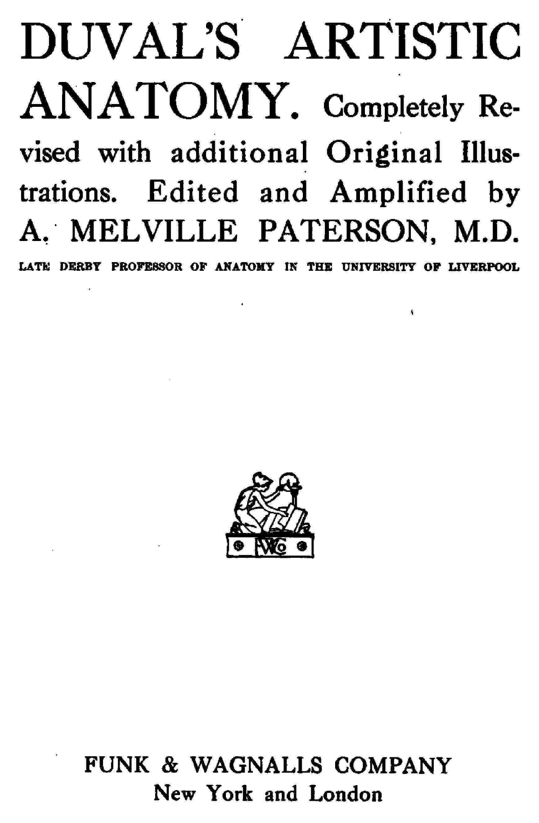
Fig. 58.
The Bony Structure of the Face.—1, the frontal bone;—2, the nasal eminence;—3, supra-orbital notch;—4, the optic foramen;—5, the sphenoidal fissure;—6, the spheno-maxillary fissure;—7, the lachrymal groove;—8, the partition and opening of the nasal fossæ;—9, the infra-orbital foramen;—10, the malar bone;—11, the symphysis of the jaw;—12, the mental foramen;—13, the ramus of the lower jaw;—14, the parietal bone;—15, the coronal suture;—16, the temporal bone;—17, the temporo-parietal suture;—18, the great wing of the sphenoid;—19, the origin of the temporal ridge;—20, the zygomatic arch;—21, the mastoid process.
The superior border is formed by the supra-orbital arch of the frontal bone (3, Fig. 58) previously described, which presents the supra-orbital notch; the inferior border is formed by the superior maxillary and malar bones; a little below its centre is a hole, called the infra-orbital foramen (9, Fig. 58), which is placed in the upper jaw almost in the same vertical line as the supra-orbital notch above the orbit (page 168). The internal border is formed by the junction of the internal angular process of the frontal bone with the ascending (nasal) process of the superior maxilla (Figs. 56 and 58). Behind the inferior part of this border is a deep groove, called the naso-lachrymal groove (7, Fig. 58), for the accommodation of the lachrymal sac (for the tears), which is the commencement of a canal (naso-lachrymal canal) connecting the orbit with the corresponding nasal fossa, and serving to transmit the nasal duct. Finally, the external border is formed by the junction of the external angular process of the frontal with the superior process of the malar bone, or bone of the cheek (10, Fig. 58; 14 and 15, Fig. 56).
The cavity of the orbit has for its walls the osseous plates belonging to the frontal (superior wall) and the several bones of the face we have previously mentioned when describing the orbital opening. We need not enter here into the description of these surfaces and of the several special bones which compose them. We shall only say that the internal wall is directed from before backwards, while the external wall is oblique, from behind forwards and from without inwards. We note, lastly, at the deepest part (towards the apex) of the cavity, three apertures, by which the orbit communicates with deeper cavities; first, a circular orifice called the optic foramen (4, Fig. 58); then, on the outer side of these, two fissures directed outwards, one obliquely upwards (sphenoidal fissure, 5, Fig. 58), and the other obliquely downwards (spheno-maxillary fissure, 6, Fig. 58). The two first communicate with the cranial cavity; the last-named fissure leads into the spheno-maxillary fossa.
The orifice of the nasal fossæ (8, Fig. 58) is situated in the middle of the face below the level of the orbits. It is somewhat heart-shaped (with the base downwards); it is bounded below by the two superior maxillary bones which unite in the middle line and form the anterior nasal spine, upon the sides by the same bones, and above by the two small nasal bones (Fig. 56, page 165) wedged in on each side of the middle line between the nasal processes of the maxillary bones, and articulating above with the nasal notch of the frontal bone.
Below, and to the outer side of each orbit, is the prominence of the cheek formed by the malar bone (10, Fig. 58). This bone is formed like a star with four rays. The superior or orbital process (15, Fig. 56) joins the external angular process of the frontal bone; the anterior process (17, Fig. 56) forms with the superior maxillary the inferior boundary of the orbit; the posterior or zygomatic process (16, Fig. 56) is directed backwards to form by its union with the zygomatic process of the temporal bone, the zygomatic arch; the inferior angle is reduced to a prominent margin which joins with the body of the bone to form the prominence of the cheek. This prominence is due to the projection of a malar process of the upper jaw on which the malar bone is fitted.
There remain now only two bones to examine on the skeleton of the face—namely, the two bones which bound the cavity of the mouth and support the teeth: these are the superior and inferior maxillary or jawbones. The superior maxillary bone (18, Fig. 56) has been in a great measure described already in relation to the orbital and nasal openings. The following points remain to be noticed: 1. The external surface is divided by a ridge descending from the malar process, into two surfaces—one outer, belonging to the zygomatic fossa, and the other anterior, appearing on the face. The facial surface is again subdivided into two smaller fossæ—the canine fossa, in which occurs the infra-orbital foramen, and the incisive fossa, above the incisor teeth—by a ridge (the canine ridge) which is formed by the prominent fang of the canine tooth. 2. The inferior or alveolar border presents a series of cavities for the lodgment of the roots of the upper teeth. The presence of these cavities is marked on the surface of the alveolar border by a series of prominences separated by depressions corresponding to the partitions between the alveoli.
Fig. 59.
The Inferior Maxillary Bone (side view).—1, the body of the inferior maxillary bone and its external oblique line;—2, the ramus;—3, the symphysis of the chin;—4, the mental foramen;—8, the angle of the jaw;—10, coronoid process;—11, the condyle;—12, sigmoid notch;—I, incisor teeth;—c, canine tooth;—b, bicuspid;—m, molar teeth.
The inferior maxillary bone or mandible (Fig. 59) requires more detailed consideration than the other bones of the face, as it takes so direct a share in the surface form that we may say that all the details of its shape are marked in the configuration of the chin and lower parts of the cheeks. It is originally composed of two distinct halves, one right and one left, which are joined together in early life in the middle line of the chin to form the symphysis of the chin, or mental process (11, Fig. 58). It is sufficient to describe one of these halves as we see it in a side view of the skull (Fig. 56).
We see that each half is formed of two strong osseous plates joined together at an angle more or less approaching a right angle, of which the prominence directed downwards and backwards is called the angle of the jaw. The horizontal portion of the bone is called the body; the vertical portion is the ramus.
The ramus consists of a flat external surface, which is continued below into the body of the bone (2, Fig. 59). Its posterior border is thick, and is continued downwards to the angle of the jaw. The anterior border, which is thinner, is grooved, and terminates by joining below the upper alveolar border of the body of the bone. The borders of the groove join the oblique lines on the surfaces of the body of the bone. The superior border is divided by a deep notch (sigmoid notch, 12, Fig. 59) into two very prominent parts. The posterior prominence is thick, and forms the neck, surmounted by the articular head, or condyle, of the jaw (11, Fig. 59), for articulation with the temporal bone; the anterior prominence is in the form of a triangular plate, and bears the name of the coronoid process (10, Fig. 59); it gives insertion to the temporal and masseter muscles.
The body of the lower jaw extends from the angle to the symphysis of the chin (3, Fig. 59); it has an external surface marked by an oblique line, above which is an orifice (12, Fig. 58; and 4, Fig. 59) called the mental foramen, placed in the same vertical line as the supra-orbital notch and the infra-orbital foramen. It is situated about the level of the second premolar tooth. The inferior border of the bone is thick and rounded; its superior alveolar border presents sockets for the teeth, and externally a series of prominences and depressions corresponding to the alveoli and the intervals between them.
The character of the lower jaw changes with age; in the infant, the angle is very obtuse and but slightly prominent: in the adult it becomes almost a right angle: in the aged the form of the jaw is changed by the loss of the teeth and by the absorption of the alveolar border, causing a diminution of height in the body of the bone. In order, therefore, to bring the two jaws in contact with one another, the lower jaw requires to move strongly forwards and upwards, whence occurs a characteristic prominence of the chin in the aged, which seems to project upwards and forwards to meet the prominence of the nose.
The teeth of the adult are altogether thirty-two in number—eight in each lateral half of each jaw. Each tooth is composed of a part fixed in the alveolar cavity called the root, and a free part called the crown. The form of the crown permits the division of the teeth into four distinct classes, which in each half of the jaw are arranged in the following manner, beginning from the median line:—Two incisors (I, Fig. 59), one canine (c, Fig. 59), two premolars, or bicuspids (b, Fig. 59), and three molars (m, Fig. 59)—total, eight. Situated at the most external and posterior portion of the dental arch, the premolar and molar teeth are hidden by the cheeks, and we need only mention that they are characterised by a crown formed of numerous tubercles (two for the premolars, four or five for the molar teeth). On the other hand, the canine and incisor teeth are easily seen when the lips are separated. The canine teeth are characterised by a conical crown with a sharp extremity, which is very large and prominent in the carnivora—e.g., in the dog (whence the name of canines). The incisors present a crown flattened from before backwards, and rectangular in form (square). Their relative size is so constant that it should be stated here. The two largest are the median incisors of the upper jaw; next in order come the lateral incisors of the upper jaw, then the lower lateral, and finally the lower median incisors, which are the smallest. The lower incisors, besides being smaller, are characterised by their chisel-like cutting edge, which is bevelled at the expense of the outer surface.
The articulation of the lower jaw with the skull, or temporo-maxillary articulation, is formed by the articulation of the condyle of the jaw (11, Fig. 59; and 29, Fig. 56) with the glenoid cavity of the temporal bone—a cavity placed in front of the external auditory meatus, and behind the articular eminence—the transverse root of the zygomatic process (page 169). This glenoid cavity, together with the articular eminence, is lined with cartilage, and is separated from the condyle of the jaw by an inter-articular fibro-cartilage. These structures are enclosed in a fibrous capsule which surrounds the articulation, and is strengthened on the outer side by a stout external lateral ligament, attached obliquely from a tubercle at the root of the zygoma downwards and backwards to the outer side of the condyle of the jaw. Therefore, when the jaw is depressed by a movement of rotation of the mandibular condyle upon its axis, this external lateral ligament is made tense, and draws the condyle forward, causing it to leave the glenoid cavity and come in contact with the articular eminence. Thus, when the mouth is widely opened (the lower jaw being greatly depressed) there is a displacement of the condyle of the jaw forwards, which is easily seen in thin subjects, and which should be noted here with its own particular mechanism.
The face, as a whole, presents a special interest when we compare its configuration with that of the cranium, in various individuals and races. In general, the more prominent the skeleton of the face the less the cranium (the forehead) is developed. This was the idea of Camper, a Dutch anatomist and artist, about the middle of the eighteenth century.
Fig. 60.
The Measurement of the Facial Angle (goniometer applied to a skull).—1, the inferior horizontal plane of the goniometer;—2, movable piece with a pin introduced into the auditory meatus;—4, graduated circle;—5, the oblique plane attached below by a hinge to the horizontal;—6, the rack for placing the bar (7) on the prominent part of the forehead.
Fig. 61.
The Facial Angle of a Skull of the Caucasian Race (after Camper).—a b and c d, the lines which mark this angle (see the text);—1, the auditory meatus;—2, anterior nasal spine;—3, the most prominent part of the forehead.
Camper proposed to measure the relative proportions of the cranium and face by the angle which the plane of the profile of the face makes with that of the base of the skull. This facial angle has since been the subject of much study on the part of anatomists and anthropologists, who have modified and perfected the process of measurement. It will be sufficient here to show what Camper’s idea was, and that, apart from anatomical considerations, he designed to furnish artists with a means of giving character to the different physiognomies of men and animals. This angle is determined by two planes (upon a head seen in profile, by two lines): one plane, which we may call horizontal, proceeds from the external auditory meatus to the anterior nasal spine, and corresponds to the inferior border of the orifice of the nasal fossa (1, Fig. 60; and a, b, Fig. 61); the other, directed obliquely upwards and backwards, is at a tangent below to the prominence of the incisor teeth, and above to the most prominent part of the forehead (c, d, Fig. 61). Fig. 60 gives an idea of the apparatus with which we measure the facial angle at the present day. It represents the facial goniometer of Jacquart. The mode of measurement here differs from that employed by Camper, in that the inferior or horizontal plane passes forward not by the nasal spine, but by the prominence of the incisor teeth.
Figures 61 and 62, which are reproduced from those of Camper, show on the one hand that while the facial angle is never equal to a right angle, it approaches to it in the best types of the white race.
62.
The Facial Angle of a Negro (after Camper). The figures are the same as in the preceding.
The ancients sought by an exaggeration to idealise the profile of the human face, and by increasing the fulness of the forehead they have given to heads of gods and heroes a facial angle as large as ninety degrees (Fig. 63). These figures show, also, the decrease of the facial angle in proportion as we pass from the white to the yellow and black races:—“The angle which the facial line or characteristic line of the visage makes,” said Camper, “varies from seventy to eighty degrees in the human species. All who raise it higher disobey the rules of art (from imitation of the antique); all who bring it lower fall into the likeness of the monkeys. If I cause the facial line to fall in front I have an antique head; if I incline it backwards I have the head of a negro; if I cause it to incline still further I have the head of a monkey; inclined still more, I have that of a dog; and, lastly, that of a goose.”[6] The figures which explain these ideas are as follows:—The facial angle of Camper averages 80 degrees in the Caucasian race; 75 degrees in the yellow, or Mongol; 60 to 70 degrees in the Negro; 31 degrees in the great monkeys (gorilla); lastly, 25 degrees in the head of a Newfoundland dog.
Fig. 63.
The Facial Angle of an Antique Head (Apollo Belvedere)—(Camper).
In our study of the various segments of the limbs we have seen that some of them have been chosen, in different systems of measurement, to serve as a common measure for these limbs, and for the entire body. Thus we have spoken of the canons which respectively take as a unit the hand (contained about ten times in the height of the body), the foot (contained a little more than six times in the total height), the middle finger (contained nineteen times), &c., &c. It is true, also, that the head—i.e., the vertical distance from its summit to the base of the chin—may be taken also as a common measure. This was done long ago. Vitruvius, speaking of the proportions of the human body, states that the height of the head should be the eighth part of the whole body. Leonardo da Vinci, Albert Dürer, and J. Cousin have followed the rule of the Latin author; and the law which makes the head the eighth of the total height has for a long time past become classic in all the schools. The choice of the head as a unit seems sufficiently justified by the two facts that, on the one hand, in every representation of the human body the head is always visible, and forms a part distinct from the rest of the body, and that, admitting that it makes the eighth part of the height, this number is particularly convenient, not being too great; and, on the other hand, it is divisible by two. In this respect it offers, for example, a great advantage over that of nineteen, which represents the proportion of the middle finger to the height.
Fig. 64.
The Facial Angle of a Monkey.
(Camper.)
Gerdy, who has adopted the law of eight heads, divides the height of the body in the following way: the first division comprises the head itself; the second extends from the chin to the level of the nipples; the third from the nipples to the umbilicus; the fourth from the umbilicus to the symphysis pubis; the fifth from the pubis to the middle of the thigh; the sixth from the thigh to the knee; the seventh from the knee to the middle of the leg; and, lastly, the eighth, from the middle of the leg to the sole of the foot (Fig. 65).
Fig. 65.
Outline of the Human Body (and proportions).
The face itself can further be subdivided into subordinate parts. The classical method of subdivision is to say that the head is the length of four noses: one from the top of the head to the top of the forehead (hair), one each for the forehead and nose, and one for the part below the nose. This is generally, however, incorrect. It is better to divide the face into two portions across the equators of the eyeballs; and subdivide the lower half into two, for the greater part of the nose above, and the lips, mouth, and chin below.
Now, if we submit to experiment the system of the law of eight heads, we see that it is accurate only in subjects of great height—for those who attain seventy-four inches and over; below seventy-two inches the subjects do not measure more than seven and a half, or only seven times the height of their head. In fact, the height of the head is a quantity which varies very little according to the subject; it is on the average, as an absolute measurement, from 8⅔ to 9 inches, and the variations which this value may present do not range below 8¼ inches or above 9 inches. A subject who measures eight heads is very tall (9 × 8 = 72, equal, or superior, to 72 inches); and a subject who only measures seven heads is of short stature (8⅔ × 7 = 61, equal, or more frequently exceeding, 61 inches).
This difference in the number of heads that the body measures in relation to absolute height seems more interesting than the narrow theory which would assign strictly the length of eight heads to each human figure. This absolute system does not agree with that which observation proves correct. Besides, it would be an error to suppose that the ancient sculptors would be slaves to such a system of proportions, since we find in their works precisely the same variations that we do in nature. The Gladiator, it is true, measures eight heads; but at the first glance at this chef d’œuvre we have the impression of a subject of great stature—of a man tall and spare. The Apollo and the Laocoon measure only 7⅔, and the Antinous only 7½ heads.
The variations in height are almost solely caused by the greater or lesser length of the lower limbs. Whether the subject be tall or short, the trunk (with the head and neck) varies comparatively little; but the thighs and legs make the differences of length. Regarding the diversity that we meet with in this question, we see that Gerdy himself has not been exact in indicating the points where the lengths of the head begin and terminate which divide the lower limb, the middle of the thigh, and the lower part of the knee; those points are badly defined, especially as he does not indicate precisely the superior extremity of the thigh. But the looseness and contradiction becomes still more evident when we come to seek, according to the various authors, the intersection between the fourth and fifth head; that is to say, the middle of the body. Without speaking of the singular inconsistency of Vitruvius, who places the middle of the body at the level of the navel, we will note only this fact, that for the passage from the fourth head to the fifth some take the pubis, others some other point.
The centre of height falls lower as the stature of the subject is increased. In subjects of small stature the centre of height corresponds to the symphysis of the pubis; for those of middle height and over, it falls about half an inch below the pubis. But it may be situated at a still lower level, and the artists of antiquity have frequently placed it much lower. In fact, as Professor Sappey says, the taller the stature, the more the centre of the body tends to fall below the symphysis, and the figures of heroes and gods are of tall stature.
We will say, then, in conclusion: 1, that the head, compared with the height, is shorter as the height increases; 2, that to produce a human figure, the absolute dimensions of which would give the impression of a subject of short stature, it would be necessary to give it about 7½ heads, and to cause the centre of the body to fall on the symphysis pubis; while to produce a figure to give the impression of tall stature it would be necessary to give it 8 heads, and to place the centre of the body more or less below the symphysis pubis.