CHAPTER X.
In order to minimise repetition and digression the following account of the Calamarieae is divided into sections, under each of which a certain part of the subject is more particularly dealt with. After a brief sketch of the history of our knowledge of Calamites, and a short description of the characteristics of the genus, the morphological features are more fully considered. A description of the most striking features of the better known Calamitean types is followed by a short discussion on the question of nomenclature and classification, and reference is made to the manner of occurrence of Calamites and to some of the possible sources of error in identification.
D. Calamites.
I. Historical Sketch.
In the following account of the Calamarieae the generic name Calamites is used in a somewhat comprehensive sense. As previous writers have pointed out, it is probable that under this generic name there may be included more than one type of plant worthy of generic designation. Owing to the various opinions which have been held by different authors, as to the relationship and botanical position of plants now generally included in the Calamarieae, there has been no little confusion in nomenclature. Facts as to the nature of the genus Calamites have occasionally to be selected from writings containing many speculative and erroneous views, but the data at our disposal enable us to give a fairly complete account of the morphology of this Palaeozoic plant.
In the earliest works on fossil plants we find several figures of Calamites, which are in most cases described as those of fossil reeds or grasses. The Herbarium diluvianum of Scheuchzer[580] contains a figure of a Calamitean cast which is described as probably a reed. Another specimen is figured by Volkmann[581] in his Silesia subterranea and compared with a piece of sugar-cane. A similar flattened cast in the old Woodwardian collection at Cambridge is described by Woodward[582] as “part of a broad long flat leaf, appearing to be of some Iris, or rather an Aloe, but ’tis striated without.” Schulze[583], one of the earlier German writers, figured a Calamitean branch bearing verticils of leaves, and described the fossil as probably the impression of an Equisetaceous plant. It has been pointed out by another German writer that the Equisetaceous character of Calamites was recognised by laymen many years before specialists shared this view.
One of the most interesting and important of all the older records of Calamites is that published by Suckow[584] in 1784. Suckow is usually quoted as the author of the generic name Calamites; he does not attempt any diagnosis of the plant, but merely speaks of the specimens he is describing as “Calamiten.” The examples figured in this classic paper are characteristic casts from the Coal-Measures of Western Germany. Suckow describes them as ribbed stems, which were found in an oblique position in the strata and termed by the workmen Jupiter’s nails (“Nägel”). Previous writers had regarded the fossils as casts of reeds, but Suckow correctly points out that the ribbed character is hardly consistent with the view that the casts are those of reeds or grasses. He goes on to say that the material filling up the hollow pith of a reed would not have impressed upon it a number of ribs and grooves such as occur on the Calamites. He considers it more probable that the casts are those of some well-developed tree, probably a foreign plant. Equisetum giganteum L. is mentioned as a species with which Calamites may be compared, although the stem of the Palaeozoic genus was much larger than that of the recent Horse-tail. The tree of which the Calamites are the casts must, he adds, have possessed a ribbed stem, and the bark must also have been marked by vertical ribs and grooves on its inner face. It is clear, therefore, that Suckow inclined to the view that Calamites should be regarded as an internal cast of a woody plant. Such an interpretation of the fossils was generally accepted by palaeobotanists only a comparatively few years ago, and the first suggestion of this view is usually attributed to Germar, Dawes, and other authors who wrote more than fifty years later than Suckow.
One of the earliest notices of Calamites in the present century is by Steinhauer[585], who published a memoir in the Transactions of the American Philosophical Society in 1818 on Fossil reliquia of unknown vegetables in the Carboniferous rocks. He gives some good figures of Calamitean casts under the generic name of Phytolithus, one of those general terms often used by the older writers on fossils. Among English authors, Martin[586] may be mentioned as figuring casts of Calamites, which he describes as probably grass stems. By far the best of the earlier figures are those by Artis[587] in his Antediluvian Phytology. This writer does not discuss the botanical nature of the specimens beyond a brief reference to the views of earlier authors. Adolphe Brongniart[588], writing in 1822, expresses the opinion that the Calamites are related to the genus Equisetum, and refers to M. de Candolle as having first suggested this view. In a later work Brongniart[589] includes species of Calamites as figured by Suckow, Schlotheim, Sternberg and Artis in the family Equisetaceae. Lindley and Hutton[590] give several figures of Calamites in their Fossil flora, but do not commit themselves to an Equisetaceous affinity.
An important advance was made in 1835 by Cotta[591], a German writer, who gave a short account of the internal structure of some Calamite stems, which he referred to a new genus Calamitea. The British Museum collection includes some silicified fragments of the stems figured and described by Cotta in his Dendrolithen. Some of the specimens described by this author as examples of Calamitea have since been recognised as members of another family.
In 1840 Unger[592] published a note on the structure and affinities of Calamites, and expressed his belief in the close relationship of the Palaeozoic plant and recent Horse-tails.
An important contribution to our knowledge of Calamites was supplied by Petzholdt[593] in 1841. His main contention was the Equisetaceous character of this Palaeozoic genus. The external resemblance between Calamite casts and Equisetum stems had long been recognised, but after Cotta’s account of the internal structure it was believed that the apparent relation between Equisetum and Calamites was not confirmed by the facts of anatomy. Petzholdt based his conclusions on certain partially preserved Permian stems from Plauenscher Grund, near Dresden. Although his account of the fossils is not accurate his general conclusions are correct. The specimens described by Petzholdt differ from the common Calamite casts in having some carbonised remnants of cortical and woody tissue. A transverse section of one of the Plauenscher Grund fossils is shown in fig. 70. The irregular black patches were described by Petzholdt as portions of cortical tissue, while he regarded the spaces as marking the position of canals like the vallecular canals in an Equisetum. Our more complete knowledge of the structure of a Calamite stem enables us to correlate the patches in which no tissue has been preserved with the broad medullary rays, which separated the wedge-shaped groups of xylem elements; the latter being more resistant were converted into a black coaly substance, while the cells of the medullary rays left little or no trace in the sandstone matrix. The thin black line, which forms the limit of the drawing in fig. 70, external to the carbonised wood, no doubt marks the limit of the cortex, and the appendage indicated in the lower part of the figure may possibly be an adventitious root. It is interesting to note that Unger[594] in 1844 expressed the opinion, which we now know to be correct, that the coaly mass in the specimens described by Petzholdt represented the wood, and that there was no proof of the existence of canals in the cortex as Petzholdt believed.
Turning to Brongniart’s later work[595] we find an important proposal which led to no little controversy. While retaining the genus Calamites for such specimens as possess a thin bark and a ribbed external surface, showing occasional branch-scars at the nodes, and having such characters as warrant their inclusion in the Equisetaceae, he proposes a second generic name for other specimens which had hitherto been included in Calamites. The fossils assigned to his new genus Calamodendron are described as having a thick woody stem, and as differing from Equisetum in their arborescent nature. Brongniart’s genus Calamodendron is made to include the plants for which Cotta instituted the name Calamitea, and it is placed among the Gymnosperms. This distinction between the Vascular Cryptogam Calamites and the supposed Gymnosperm Calamodendron is based on the presence of secondary wood in the latter type of stem. The prominence formerly assigned to the power of secondary thickening possessed by a plant as a taxonomic feature, is now known to have been the result of imperfect knowledge. The occurrence of a cambium layer and the ability of a plant to increase in girth by the activity of a definite meristem, is a feature which some recent Vascular Cryptogams[596] share with the higher plants; and in former ages many of the Pteridophytes possessed this method of growth in a striking degree.
Although Brongniart’s distinction between Calamites and Calamodendron has not been borne out by subsequent researches, the latter term is still used as a convenient designation for a special type of Calamitean structure. One of the earliest accounts of the anatomy of Calamodendron stems is by Mougeot[597], who published figures and descriptions of two species, Calamodendron striatum and C. bistriatum.
Some years later Göppert[598], who was one of the greatest of the older palaeobotanists, instituted another genus, Arthropitys[599], for certain specimens of silicified stems from the Permian rocks of Chemnitz in Saxony, which Cotta had previously placed in his genus Calamitea under the name of Calamitea bistriata[600]. Göppert rightly decided that the plants so named by Cotta differed in important histological characters from other species of Calamitea. The generic name Arthropitys has been widely adopted for a type of Calamitean stem characterised by definite structural features. The great majority of the petrified Calamite stems found in the English Coal-Measures belong to Göppert’s Arthropitys.
The next proposal to be noticed is one by Williamson[601] in 1868; he instituted the generic name Calamopitys for a few examples of English stems, which differed in the structure of the wood and primary medullary rays from previously recorded types. We have thus four names which all stand for generic types of Calamitean stems. Of these Calamodendron and Arthropitys are still used as convenient designations for stems with well-defined anatomical characters. The genus Calamitea is no longer in use, and Williamson’s name Calamopitys had previously been made use of by Unger[602] for plants which do not belong to the Calamarieae. As it is convenient to have some term to apply to such stems as those which Williamson made the type of Calamopitys, the name Arthrodendron is suggested by my friend Dr Scott[603] as a substitute for Williamson’s genus.
The twofold division of the Calamites instituted by Brongniart has already been alluded to, and for many years it was generally agreed that both Pteridophytes and Gymnosperms were represented among the Palaeozoic fossils known as Calamites. The work of Prof. Williamson was largely instrumental in proving the unsound basis for this artificial separation; he insisted on the inclusion of all Calamites in the Vascular Cryptogams, irrespective of the presence or absence of secondary wood. By degrees the adherents of Brongniart’s views acknowledged the force of the English botanist’s contention. It is one of the many signs of the value of Williamson’s work that there is now almost complete accord among palaeobotanical writers as to the affinities of Calamitean plants.
In the following account of the Calamites, the generic name Calamites is used in a wide sense as including stems possessing different types of internal structure; when it is possible to recognise any of these structural types the terms Calamodendron, Arthropitys or Arthrodendron are used as subgenera. The reasons for this nomenclature are discussed in a later part of the Chapter.
| Genus Calamites, Suckow, 1714 | This term was originally applied to the common pith-casts of Calamitean stems, without reference to internal structure. |
| Subgenera | Calamodendron, | Brongniart, | 1849 | These names have primarily reference to internal structure. | |
| Arthropitys | Göppert, | 1864 | |||
| Arthrodendron | Scott | 1897 | |||
| (= Calamopitys | Williamson, | 1871 |
II. Description of the anatomy of Calamites.
No fossils are better known to collectors of Coal-Measure plants than the casts and impressions of the numerous species of Calamites. In sandstone quarries of Upper Carboniferous rocks there are frequently found cylindrical or somewhat flattened fossils, varying from one to several inches in diameter, marked on the surface by longitudinal ridges and grooves, and at more or less regular intervals by regular transverse constrictions. Similar specimens are still more abundant as flattened casts in the blocks of shale found on the rubbish heaps of collieries. The sandstone casts are often separated from the surrounding rock by a loose brown or black crumbling material, and the specimens in the shale are frequently covered by a thin layer of coal.
Most of the earlier writers regarded such specimens as the impressions of the ribbed stems of plants similar to or identical with reeds or grasses. Suckow, and afterwards Dawes and others, expressed the opinion that the ordinary Calamite cast represented a hardened mass of sand or marl, which had filled up the pith of a stem either originally fistular or rendered hollow by decay. The investigation of the internal structure confirmed this view, and proved that the surface-features of a Calamite stem do not represent the external markings of the original plant, but the form of the inner face of the cylinder of wood. The ribs represent the medullary rays of the original stem or branch, and the intervening grooves mark the position of the strands of xylem which are arranged in a ring round a large hollow pith[604].
With this brief preliminary account we may pass to a detailed description of the anatomical characters of Calamites.
The genus Calamites may be briefly defined as follows:—
Arborescent plants reaching a height of several meters, and having a diameter of proportional size. In habit of growth the Calamites bore a close resemblance to Equisetum; an underground rhizome giving off lateral branches and erect aerial shoots bearing branches, either in whorls from regularly recurring branch-bearing nodes, or two or three from each node; and in some cases the stems bore occasional branches from widely separated nodes. The leaves were disposed in whorls either as star-shaped verticils on slender foliage shoots, or in the form of a circle of long narrow leaves on the node of a thicker branch. Adventitious roots were developed from the nodal regions of underground and aerial stems. The cones had the form of long and narrow strobili consisting of a central axis bearing whorls of sterile and fertile appendages; the latter in the form of sporangiophores bearing groups of sporangia. The strobili were heterosporous in some cases, isosporous in others. The stems had a large hollow pith bridged across by a transverse diaphragm at the nodes in the centre of the single stele; the latter consisted of a ring of collateral bundles separated from one another by primary medullary rays. Each group of xylem was composed of spiral, annular, scalariform and occasionally reticulate tracheids, the position of the protoxylem being marked by a longitudinal carinal canal. The shoots and roots grew in thickness by means of a regular cambium layer. The cortex consisted of parenchymatous and sclerenchymatous cells, with scattered secretory sacs. The increase in girth of the central cylinder was often accompanied by a considerable development of cortical periderm. The roots differed from the shoots in having no carinal canals, and in the possession of a solid pith and centripetally developed primary xylem groups alternating with strands of phloem.
The above incomplete diagnosis includes only some of the more important structural features of the genus. Thanks to the researches begun by the late Mr Binney of Manchester and considerably extended by Carruthers, Williamson and later investigators, we are now in a position to give a fairly complete account of Calamites. The type of stem most frequently met with in a petrified condition in the English rocks is that to which Göppert applied the name Arthropitys, and it is this subgenus that forms the subject of the following description. Our knowledge of Calamitean anatomy is based on the examination of numerous fragments of petrified twigs and other portions of different specific types of the genus. It is seldom possible to differentiate specifically between the isolated fragments of stems and branches which are met with in calcareous or siliceous nodules. As so frequently happens in fossil-plant material, large specimens showing good surface features and broken fragments with well-preserved internal structure have to be dealt with separately.
a. Stems.
A transverse section of a young twig, such as is represented in fig. 71, illustrates the chief characteristics of the primary structure of a young branch of Calamites. The figure has been drawn from a section originally described by Hick[605] in 1894. A very young Calamite twig bears an exceedingly close resemblance to the stem of a recent Equisetum. The axial region of the stem may be occupied by parenchymatous cells, or the absence of cells in the centre may indicate the beginning of the gradual formation of the hollow pith, which is one of the characteristics of Calamites. The student of petrified Palaeozoic plants must constantly be on his guard against the possible misinterpretation of Stigmarian ‘rootlets,’ which are frequently found in intimate association with fossil tissues. The intrusion of these rootlets is admirably illustrated by a section of a Calamite stem in the Williamson Collection (No. 1558) in which the hollow pith, 2 cm. broad, contains more than a dozen Stigmarian appendages.
In the figured specimen of a Calamite twig (fig. 71) there is a clearly marked differentiation into a cortical region and a large stele or central cylinder. The pith-cells are already partially disorganised, but there still remain a few fairly large parenchymatous cells internal to the ring of vascular bundles. The few irregular projections into the cavity of the large pith consist of small fragments of cells, which may be the result of fungal action. Mycelia of fungi are occasionally met with in the tissues of older Calamite stems.
The position of the primary xylem groups is shown by the conspicuous and regularly placed canals, c; these have been formed in precisely the same manner as the corresponding spaces in an Equisetum stem, and they are spoken of in both genera as the carinal canals. Each canal owes its origin to the disorganization and tearing apart of the protoxylem elements and the surrounding cells. This may be occasionally seen in examples of very young Calamites; the canals of a young twig often contain apparently isolated rings which are coils of elongated spiral threads. Fig. 72, B represents the canal of a twig, cut in an oblique direction, in which the remains of spiral tracheids are distinctly seen. In the stem of fig. 71 the development has not advanced far enough to enable us to clearly define the exact limits of each xylem strand. The smaller elements bordering the canals constitute the primary xylem, they are fairly distinct on the outer margin of some of the canals seen in the section. Between the small patches of primary xylem the outward extensions of the parenchyma of the pith constitute the primary medullary rays, mr. The distinct line encircling the canals and primary xylem has been described by Hick as marking the position of the endodermis, but it may possibly owe its existence to the tearing of the tissues along the line where cambial activity is just beginning. This layer of delicate dividing cells would constitute a natural line of weakness. External to this line we have a zone of tissue a, d, containing here and there larger cells with black contents, which are no doubt secretory sacs. It is impossible to distinguish with certainty any definite phloem groups, but in other specimens these have been recognised immediately external to each primary xylem group; the bundles were typically collateral in structure. Towards the periphery of the twig the preservation is much less perfect; the outer portion of the inner cortex, d, consists of rather smaller and thicker-walled cells, but this is succeeded by an ill-defined zone containing a few scattered cells, b, which have been more perfectly preserved. The twig is too young to show any secondary tissue in the cortex; but the tangential walls in some of the cortical cells afford evidence of meristematic activity, which probably represents the beginning of cork-formation. The limiting line, e, possibly represents the cuticularised outer walls of an epidermal layer. The irregularly wavy character of the surface of the specimen is probably the result of shrinking, and does not indicate original surface features.
In examining sections of calcareous nodules from the coal seams one meets with numerous fragments of small Calamitean twigs with little or no secondary wood; in some of these there is a small number of carinal canals, in others the canals are much more abundant. The former probably represent the smaller ramifications of a plant, and the latter may be regarded as the young stages of branches capable of developing into stout woody shoots[606]. Longitudinal sections of small branches teach us that the xylem elements next the carinal canals are either spiral or reticulate in character, the older tracheids being for the most part of the scalariform type, with bordered pits on the radial walls. This and other histological characters are admirably shown in the illustrations accompanying Williamson and Scott’s memoir on Calamites. The student should treat the account of the anatomy of Calamites given in these pages as introductory to the much more complete description by these authors. They thus describe the course of the vascular bundles in a Calamitean branch:—
“The bundle-system of Calamites bears a general resemblance to that of Equisetum. A single leaf-trace enters the stem from each leaf, and passes vertically downwards to the next node. In the simplest cases the bundle here forks, its two branches attaching themselves to the alternating bundles which enter the stem at this node. In other cases both the forks attach themselves to the same bundle, so that, in this case, there is no regular alternation. In other cases, again, the bundle runs past one node without forking, and ultimately forms a junction with the traces of the second node below its starting-point. These variations may all occur in the same specimen. The xylem at the node usually forms a continuous ring, for where the regular dichotomous forks of the bundles are absent their place is usually taken by anastomoses[607].”
As in Equisetum, the xylem at the nodes possesses certain characteristic features which distinguish it from the internodal strands. It has already been pointed out that the xylem of Equisetum increases in breadth at the nodes (p. 251, fig. 55, 4); the same is true of Calamites. In fig. 72, C, we have part of a radial section of a Calamite twig in which the broad mass of short nodal tracheids is clearly shown; this nodal wood forms a prominent projection towards the pith. In the lower part of the section the remains of some spiral protoxylem tracheids are seen in a carinal canal.
- A. External xylem elements and cambium, c, with imperfect phloem, × 100.
- B. Carinal canal containing protoxylem, px. × 65.
- C. Radial longitudinal section through nodal xylem, px. × 35.
- D. Phloem elements; s, sieve-tubes; p, p, parenchymatous cells.
- (A–C. After Williamson and Scott. D. After Renault.)
The tracheids of the nodal wood are often reticularly pitted, and so differ in appearance from the ordinary scalariform elements.
It is rare to find the phloem clearly preserved, but in specimens where it has been possible to examine this portion of the vascular bundles, it is found to consist of elongated cambiform cells and sieve-tubes. An unusually perfect specimen has been described by Renault[608] in which the phloem elements are preserved in silica. Fig. 72, D, is copied from one of Renault’s drawings, the sieve-tubes, s, s, show several distinct sieve-plates on the lateral walls of the tubes, reminding one to some extent of the sieve-tubes in a Bracken Fern. The cells, p, p, associated with the sieve-tubes are square-ended elongated parenchymatous elements. Another characteristic feature illustrated by longitudinal sections is the nodal diaphragm; except in the smallest branches the interior of each internode is hollow, and the ring of vascular bundles is separated from the pith-cavity by a band of parenchymatous tissue. At each node this parenchyma extends across the central cavity in the form of a nodal diaphragm, as in the stem of Equisetum.
By far the greater number of the petrified fragments of Calamites afford proof of cambial activity, and possess obvious secondary tissues. In exceptionally perfect specimens the xylem tracheids are found to be succeeded externally by a few flattened thin-walled cells which are in a meristematic condition (fig. 72, A, c); these constitute the cambium zone, and it is the secondary structure that results from the activity of the meristematic cells that we have now to consider.
In petrified examples of branches in which the secondary thickening has reached a fairly advanced stage, the wood is usually the outermost tissue preserved, the more external tissues having been detached along the line of cambium cells. It is only in a few cases that we are able to examine all the tissues of older examples.
The specimen represented in fig. 73 illustrates very clearly the extension of the hollow pith up to the inner surface of the vascular ring; the disorganisation of the pith-cells which had already begun in the twig of fig. 71 has here advanced much further. The bluntly rounded projections represent the prominent primary xylem strands, each of which is traversed by the characteristic carinal canal. Alternating with the wedge-shaped groups of secondary xylem, x, we have the broad principal medullary rays, mr, which become slightly narrower towards the outside. The inner face of each of these wide rays has a concave form, due to the less resistent nature of the medullary-ray cells as compared with the stronger xylem. The regularly sinuous form of the inner face of the vascular cylinder enables one to realise how the Calamite-casts (figs. 82, 99, and 101) have come to have the regular ridges and grooves on their surface. The broad ridges on the cast mark the position of the wide medullary rays, while the grooves correspond to the more prominent ends of the vascular strands. The tissues external to the wood have not been preserved in the example shown in fig. 73. Some silicified specimens described by Stur[609] from Bohemia and now in the Museum of the Austrian Geological Survey, Vienna, admirably illustrate the connection between the surface features of a Calamite cast and the anatomy of the stem.
mr, medullary ray. After Williamson.
x, x, xylem. (No. 1933 A.A. in the Williamson Collection.)
In the large section of a calcareous nodule diagrammatically shown in fig. 17 II. (p. 85) the secondary wood of a slightly flattened Calamite is the most prominent plant fragment. The pith-cavity has been almost obliterated by the lateral compression of the woody cylinder, but the presence of the carinal canals along the inner edge of the wood may still be readily recognised. The appearance presented by a transverse section of the secondary wood of a Calamite is that of regular radial series of rather small rectangular tracheids, with occasional secondary medullary rays consisting of narrow and radially elongated parenchymatous cells. The principal rays[610] in the Arthropitys type of a Calamite stem are often found to gradually decrease in breadth as they pass into the secondary wood, until in the outer portion of the wood the primary medullary rays are practically obliterated by the formation of interfascicular xylem.
In fig. 74, A, we have a portion of a single xylem group of a thick woody stem. The stem from which the figure has been drawn was originally described by Binney[611] as Calamodendron commune; we now recognise it as a typical example of the subgenus Arthropitys. The specific term communis was used by Ettingshausen[612] in 1855 in a comprehensive sense to include more than twenty species of the genus Calamites, but since Binney’s use of the term it has come to be associated with a definite type of Arthropitys stem, in which the primary medullary rays decrease rapidly in breadth towards the periphery of the wood. The wood of Binney’s stem[613] measures 2·5 cm. across, but the pith-cavity has been crushed to the limits of a narrow band represented in the figure by the shaded portion. The strand of cells, s, in the pith is a portion of a Stigmarian appendage (“rootlet”), which penetrated into the hollow stem of the Calamite and became petrified by the same agency to which the preservation of the stem is due. These intruded Stigmarian appendages are of constant occurrence in the calcareous nodules; their intimate association with the tissues of other plants is often a serious source of error in the identification of petrified tissues. The inner portion of one of the xylem groups is shown in fig. 74, A. External to the carinal canal, the xylem tracheids are disposed in regular series and associated with numerous narrow secondary medullary rays. The width of the xylem wedge increases gradually as we pass outwards, this is due to the formation of interfascicular xylem, which in the more peripheral portion of the stem extends across the primary medullary rays. The few primary medullary-ray cells shown in the drawing illustrate the characteristic tangentially elongated form and large size of the parenchymatous elements. Williamson and Scott have pointed out that the tangentially elongated form of the medullary-ray cells is the result of active growth, and not merely the expression of the tangential stretching of the stem consequent on secondary thickening.
- Transverse section of part of a Calamite stem. [Calamites (Arthropitys) communis (Binney).]
s, Stigmarian appendage. x, xylem. From a specimen in the Binney Collection, Cambridge, × 50. - Transverse section of a stem.
h, hypodermal tissue; c, inner cortex. From a specimen in the Williamson Collection (no. 62). × 35.
A glance at the complete transverse section of the stem,—of which a small portion is shown in fig. 74 A,—suggests the existence of annual rings in the wood, but this appearance of rings is merely the result of compression. The secondary wood of a Calamite does not exhibit any regular zones of growth comparable with the annual rings of our forest trees.
x, x, secondary xylem and medullary rays; m, principal medullary ray. From a section in the Binney Collection, × 50.
Before passing to other examples of Calamitean stems, reference may be made to the sections shown in figs. 75 and 76, which illustrate some further points in the structure of Binney’s stems. In fig. 75 the xylem tracheids are shown at x, and between them the secondary medullary rays present the appearance of long and narrow parenchymatous cells; as the section is tangential the characteristic scalariform character of the tracheids is not shown, the ladder-like bordered pits being confined to the radial walls of the tracheal elements. The much greater length than breadth of the cells which form the rays associated with the xylem tracheids, is a characteristic feature in Calamitean stems. The breadth of the principal ray, m, shows that the section has passed through the wood a short distance from the pith; in a tangential section cut further into the wood the breadth of the principal rays would be considerably reduced. The large medullary-ray tissue consists of square-walled parenchymatous cells. The more highly magnified section, in fig. 76, shows a central group of parenchyma containing a few transversely cut tracheids, but the two kinds of elements are not clearly differentiated in the figure; this group of cells is an outgoing leaf-trace which is enclosed by the strongly curved tracheids of the stem. The section is taken from the node of a stem where several leaf-trace bundles are passing out to a whorl of leaves; the few cells intercalated between the tracheids belong to the parenchyma of the secondary medullary rays.
In the small portion of a stem represented in fig. 74 B, the cortical tissues have been partially preserved; at the inner edge, next the hollow pith, there are two xylem groups, each with a carinal canal, and between them is part of a broad “principal” medullary ray[614]. The cambium has not been preserved, but beyond this region we have some of the large cells, c, of the inner cortex; these are followed by a few remnants of a smaller-celled tissue, and external to this part of the cortex there is a series of triangular groups, h, consisting of small thick-walled cells alternating with spaces which were originally occupied by more delicate parenchyma. The darker groups constitute hypodermal strands of mechanical tissue or stereome which lent support to the stem. The surface of a stem possessing such supporting strands would probably assume a longitudinally wrinkled or grooved appearance on drying; the intervening parenchyma, contracting and yielding more readily, would tend to produce shallow grooves alternating with the ridges above the stereome strands.
The complete section of the stem of which a small portion is shown in fig. 74 B, is figured by Williamson[615] in his 12th memoir on Coal-Measure plants. The section was obtained from Ashton-under-Lyne in Lancashire; it illustrates very clearly a method of preservation which is occasionally met with among petrified plants. The walls of the various tissue elements are black in colour and somewhat ragged, and the general appearance of the section is similar to that of a section of a charred piece of stem. It is possible that the Calamite twig was reduced to charcoal before petrifaction by a lightning flash or some other cause.
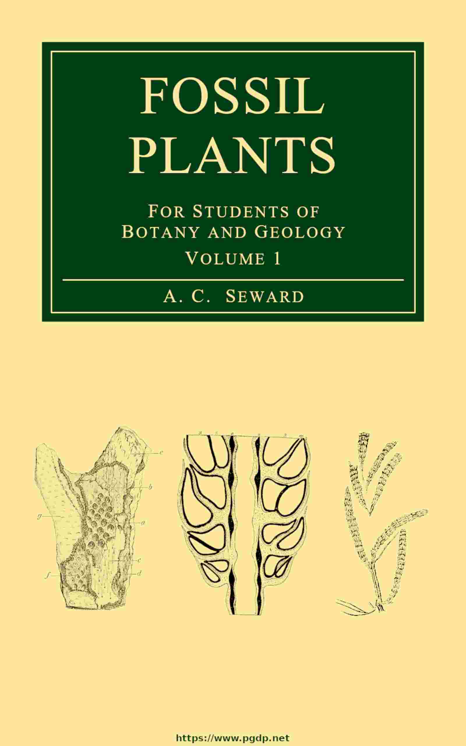
It is often said that the surface of a Calamite stem was probably marked by regular ridges and grooves similar to those of the pith-cast, and that such external features are connected with the arrangement of the tissues in the vascular cylinder. The indication of grooves and ridges on the bark of fossil Calamites is no doubt the result of the existence in the hypoderm of firm strands alternating with strands of less resistant cells. It is very common to find Calamite pith-casts covered with a layer of coal presenting a ribbed surface, but this is simply due to the moulding of the coaly film on an internal pith-cast. The broad grooves on such a specimen as that of fig. 77 are, on the other hand, probably an indication of the existence of hypoderm bands similar to those in fig. 74 B, h. The specimen from which fig. 77 is drawn shows many interesting features. The figure given by Grand’Eury, of which fig. 77 is a copy, is somewhat idealised, but the various surfaces can be made out in the fossil. The surface of the coaly envelope surrounding the pith-cast, a, is distinctly grooved, but the depressions have nothing to do with the surface features of the wood or the pith-cast; they are no doubt due to the occurrence of alternating bands of thick- and thin-walled tissue in the hypodermal region of the cortex; the peripheral strands of bast cells would stand out as prominent ribs as the stem tissue contracted during fossilisation. At b (fig. 77) we have a view of the wood in which the position of the principal rays is indicated by fine longitudinal lines at regular intervals; the oval projections just below the nodal line are probably the casts of infranodal canals (cf. p. 324). At a the characteristic pith-cast is seen with a small branch-scar on the node. The scar on the middle node, N 2, is probably that of a root, and a root R is still attached to the node, N 3.