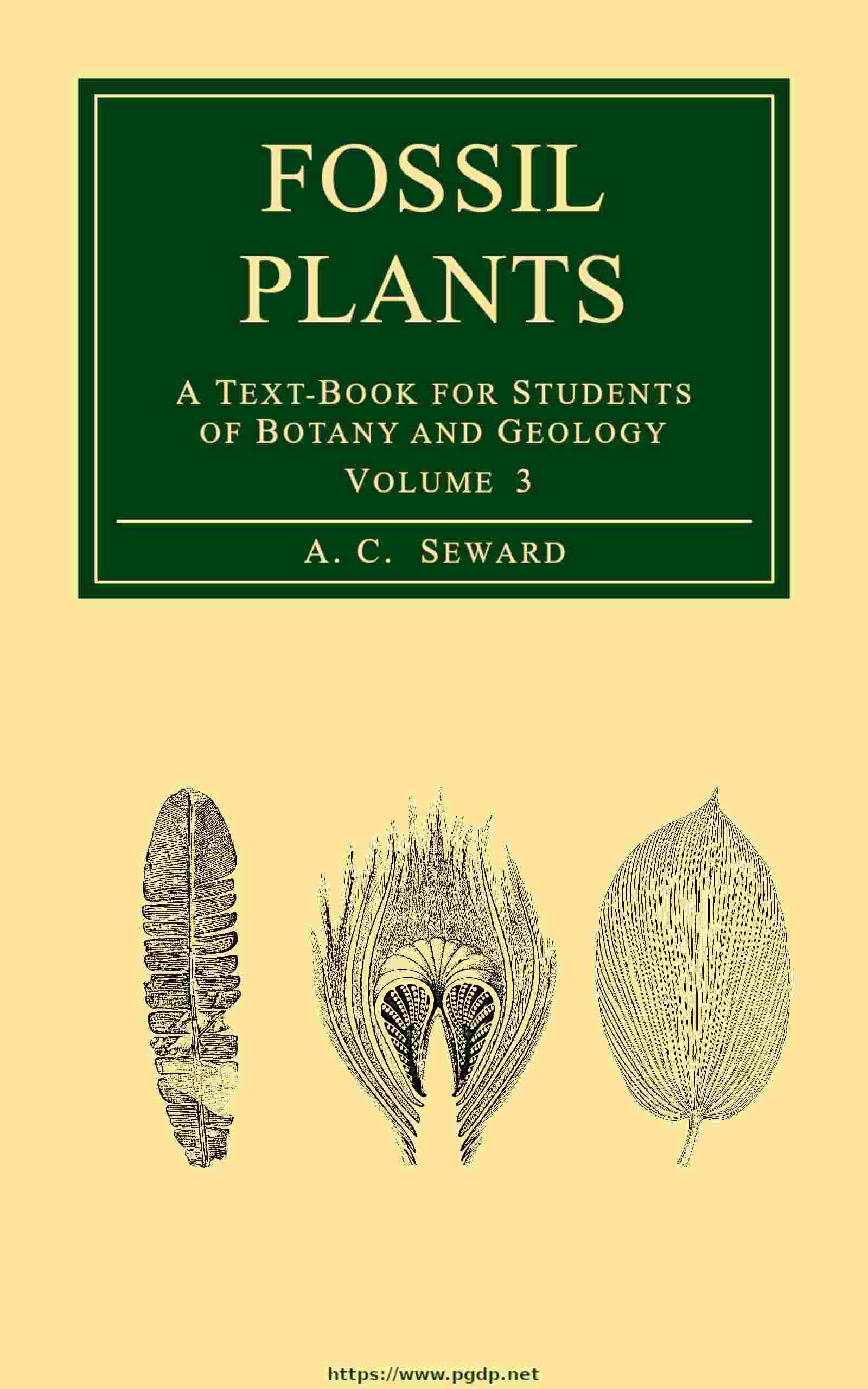
- A, B. Crossotheca Hoeninghausi.
- C. Pinnule with a sporangium, s.
Telangium. Reference was made in vol. ii.[138] to the genus Telangium instituted by Dr Benson for some petrified sporangia from the Coal Measures regarded by her as the microsporangia of a Pteridosperm, probably Lyginopteris. The sporangia of Telangium are similar to those of Crossotheca. Scott points out that they are borne on a flat disc or lamina ‘quite comparable to a fertile pinna of Crossotheca[139],’ and he concludes that these sporangia are not generically distinct from the impressions on which the genus Crossotheca was founded. Kidston[140] regards Telangium Scotti, Benson, as the microsporangium of a Pteridosperm though not of Lyginopteris, on the ground that the microsporangia described by Miss Benson are not attached to a limb and that they have a single loculus in place of the double loculus (fig. 407, A) of Crossotheca. The presence of a limb in Telangium recognised by Scott removes one of these distinguishing features. There are, however, no adequate reasons for regarding Telangium Scotti as specifically identical with Crossotheca Hoeninghausi. The synangium of Telangium Scotti, 5 mm. in length, consists of 6–12 sporangia united basally and opening when ripe by longitudinal dehiscence. Fig. 493, E, shows eight sporangia of a synangium in transverse section: the two sporangia at the lower end of the section are less distinct than the others, some are full of spores and others have shed their contents by the splitting of the thin inner walls of the loculi. The sporangial walls are composed of an outer layer of large cells with dark contents succeeded by 2–3 layers of smaller and crushed cells. The spores, 5–6 µ × 3·5–4 µ in diameter, have a reticulately sculptured exine: Dr Benson[141] states that they agree closely with pollen-grains found in the pollen-chamber of Lagenostoma ovoides except in their slightly smaller size; she notes the association of Telangium with fragments of the vegetative organs of Lyginopteris and draws attention to resemblances in the structure of the tissues; but the most interesting comparison, at least in an academic sense, is with the seed Lagenostoma, the integumented megasporangium of Lyginopteris. Dr Benson points out that a transverse section of a Lagenostoma in the plane of the canopy, showing the nucellar apex surrounded by radially disposed chambers (fig. 409), presents a certain resemblance to a synangium of Telangium Scotti; and it is suggested that the chambers encircling the nucellus may represent sterilised sister-sporangia[142]. ‘The seed in fact is assumed to be a synangium in which all but one of the sporangia are sterile, and form an integument to the one fertile sporangium which has become a megasporangium with one large megaspore.’ This view, though clearly incapable of confirmation in the present state of our knowledge, is not merely an ingenious hypothesis but a stimulating suggestion as to possible homologies: as an argument in favour of associating Lagenostoma and Telangium as the spore-bearing organs of the same plant it has but little weight.
iv. The Seed. Lagenostoma Williamson.
Lagenostoma Lomaxi, Oliver and Scott ex Williamson, MS.
In 1877 Williamson[143] proposed the generic name Lagenostoma for some petrified seeds from the Lower Coal Measures of Lancashire and described two species, Lagenostoma ovoides and L. physoides: in his MS. Catalogue a third type is referred to as Lagenostoma Lomaxi. It is this third type that Prof. Oliver was the first to recognise as the megaspore-bearing organ of Lyginopteris oldhamia. Its structure has been thoroughly described by Oliver and Scott[144] and these authors contribute a judicial summary of the evidence on which Lagenostoma and Lyginopteris are believed to stand for one and the same plant. The evidence is based chiefly on the following considerations: an agreement in the structure of the vascular bundles in the investments of the seed with those in the leaves of Lyginopteris; the presence in the outer envelope of the seed of stalked glands identical with those on the stems and petioles. The evidence does not as yet amount to absolute proof, as the seeds, which occur either with or without a stalk, have not been found attached to a Lyginopteris frond. But ‘where vegetative and reproductive organs presenting identical structural features, not known to occur in other plants, are thus found in close and constant association, the inference that the one belonged to the other appears irresistible.’ While most botanists believe that a satisfactory case is established there are a few[145] who refuse to believe in a connexion between Lagenostoma and Lyginopteris until an actual union has been demonstrated. The discovery by Kidston[146] of seeds attached to pinnae bearing Neuropteris pinnules and the demonstration of organic continuity between seeds and the pinnules of other Palaeozoic fern-like fronds supply abundant confirmatory evidence that leaves no doubt as to the occurrence of seeds on modified pinnae of Sphenopteris Hoeninghausi and of other closely allied fronds which represent the foliage of different forms of Lyginopteris. In this connexion it is pertinent to add that Grand’Eury[147] has found seeds of the Lagenostoma type in close association with impressions of Sphenopteris Dubuissonis and other leaves of similar habit.
A seed of Lagenostoma Lomaxi reaches a length of 5·5 mm. with a maximum diameter of 4·4 mm.; it is broadly oval or barrel-like (fig. 408, C) and when immature was invested by a loose irregularly lobed glandular envelope (fig. 408, B) from which the seed eventually freed itself by a natural process of abscission. The central body or nucellus, except in the apical region, is concrescent with a fairly stout integument or testa (fig. 408, C, f) the outer portion of which is characterised by regular longitudinal rows of palisade-like cells comparable with the broad palisade-layer in the sporocarp of Pilularia. On the exposed surface of this palisade-tissue are small dark structureless pegs[148], possibly the remains of a mucilaginous layer such as occurs on the seed-coats of some recent Flowering plants. At the base of the nucellus the chalazal region, fig. 408, C, ch, is provided with sclerous elements and forms a hard investment to the axial vascular strand from the pedicel. It is at the base of this chalazal region that the seed is eventually cut off by an absciss-layer. The integument is supplied throughout its length by nine vascular bundles of endarch, or approximately endarch, structure. The free portion of the integument seen from the outside (fig. 408, B) has the form of a fluted cone with a circular opening at its summit. The greater part of this domical apex, as seen in longitudinal section in fig. 408, C, appears to be hollow, but in the living state the dome, or canopy as Williamson called it, was filled with parenchyma in which the vascular bundles were embedded and, as shown in the transverse section in fig. 409, the canopy is divided into compartments by radial septa which in its basal region are replaced by regular and deep furrows on the inner face. Enclosed by the canopy, with its outer surface fluted as the result of the partial collapse of the outer wall of each compartment due to the decay of the filling tissue, is the flask-shaped apex of the nucellus; between the apical cone of nucellar parenchyma and the superficial layer is an annular cavity which Williamson[149] called the lagenostome. The parenchymatous core tapers to a narrow summit which slightly overtops the integument and is constricted at the broad base (fig. 493, A, B; page 311). The bottle-shaped apical tissue is separated by an annular space, c, fig. 493, B, from the limiting layer of the nucellus: this space is the pollen-chamber formed in the living seed by the disorganisation of the subepidermal cells of the nucellar apex. The pollen-chamber is a feature characteristic of recent cycadean ovules (see p. 6). In Lagenostoma the annular form of the pollen-chamber is a peculiarity distinguishing this type of seed from those of recent Gymnosperms and most other Palaeozoic seeds. As Oliver says, it marks an ‘advance in precision’[150] over other forms as the microspores which fall into the chamber are brought direct to the surface of the underlying megaspore and presumably to the archegonia which, it is reasonable to believe, were disposed in a circle at the base of the annular crevice. Microspores frequently occur in the pollen-chamber and some have been discovered apparently in the act of liberating male gametes[151].
The outer wall of the nucellus is bounded externally by a similar circular space (d, figs. 408, C; 493, B) which separates it from the domical canopy. In the great majority of specimens the central tissue of the seed is not preserved and an empty sac supported from the base of the nucellus-apex occupies nearly the whole of the interior: the shrunken wall of the sac is all that remains of the large megaspore. It would seem, then, that the nucellus was almost completely destroyed as a consequence of the growth of the megaspore or embryo-sac, which eventually occupied nearly the whole of the seed-body.
In an exceptionally well preserved specimen recently described and admirably illustrated by Mr McLean[152] part of the parenchymatous tissue of the prothallus which originally filled the megaspore is clearly seen: its surface-layer consists of small cells succeeded by a broad band of radially elongated elements closely resembling the alveoli in the prothalli of some recent Gymnosperms, particularly certain Conifers. No archegonia have been discovered. The cupular envelope of immature seeds, compared by Oliver and Scott with the lobed and glandular husk of Corylus colurna L.[153], receives several vascular bundles of collateral and mesarch structure from the axial strand, and these subdivide as they ascend. The glands which occur on all parts of the cupule are sessile or stalked and identical with those on the vegetative organs of Lyginopteris. Assuming that pollination occurred at a comparatively early stage in the development of the seed when the cupule was still intact, it is conceivable, as Sir Joseph Hooker suggested, that the glandular secretion may have attracted insects and so aided in the transport of pollen which were perhaps drawn down the narrow pollen-chamber by exuded mucilage as in recent Conifers. The evidence obtained in recent years in favour of insect-pollination in certain Cycads and in Welwitschia lends support to this view: the dragon flies hovering over a fertile Lyginopteris frond in a recent restoration[154] may be a legitimate addition.
A striking feature of Lagenostoma as of other Palaeozoic seeds is the absence of an embryo: this and other considerations have led certain authors, notably Chodat[155], to question the justification for the use of the term seed. Various suggestions have been offered in explanation of this fact. In recent Cycads, as already pointed out, the development of the embryo does not always occur before seed-fall. It may be that these older seeds had no resting-period or there may have been a period of rest after fertilisation and not as now at a stage subsequent to the formation of the embryo[156]; it is also suggested by Scott that ‘the nursing of the embryo had not yet come to be one of the functions of the seed, and that the whole embryonic development was relegated to the germination stage[157].’ In this connexion reference may be made to a statement by Miss Gibbs[158] who speaks of seeds of a Podocarpus picked up from the ground in apparently a mature state and with the associated bracts coloured and swollen as though ready to aid in dispersal but with no embryo: the seeds had matured before fertilisation and fell from the tree after pollination. Whatever may be the true explanation of the absence of embryos this negative character should not be allowed to outweigh the evidence furnished by morphological features as to the applicability of the term seed. As Prof. Oliver says, ‘there is a long chapter in evolution to be deciphered before we can connect ... the seed of Lyginodendron with the sporangium of any fern at present known to us’[159].
The cupule of Lagenostoma has been homologised with the outer part of the integument of a recent cycadean seed[160] which, it is suggested, consists of an inner and an outer envelope that have become concrescent, and this hypothesis is supported by another author by a comparison between Lagenostoma and Gnetalean seeds[161]. A comparison has also been made between the collar of a Ginkgo seed and the much more conspicuous cupule of Lagenostoma[162]. Dr Benson and Miss Welsford[163] institute a comparison between the vascular supply of the outer integument of the ovules of Carpinus and Morus and that of the cupule of Lagenostoma, a comparison suggested by Miss Kershaw’s remarks[164] on the similarity between the vascular system of the ovules of Myrica Gale and Trigonocarpus. In 1908 Dr Benson[165] described some germinating microspores in the pollen-chamber of another species of Lagenostoma, L. ovoides, and recognised what she believed to be antherozoids. I am indebted to this author for allowing me to make a drawing from her section (fig. 408, D). Two microspores are seen with thick outer walls showing irregular holes probably of secondary origin and not part of a regular reticulum as Dr Benson suggests. Close to the upper microspore is a hemispherical body, s, described as a male gamete, and a similar body is seen still enclosed by the lower microspore. It is by no means improbable that these are antherozoids: they were presumably ciliate like those of Ginkgo and recent Cycads (fig. 396, M). The microspores are approximately 70μ in length and the supposed antherozoids have a maximum diameter of 45μ, the latter being about ⅔ the size of the sperms of Microcycas and ⅙ the diameter of those of Zamia. The smaller and more delicate cells near the lower microspore (fig. 408, D) are no doubt fungal cells as Miss Benson suggests. With reference to the difficulty of determining the nature of Miss Benson’s supposed gametes it is worth calling attention to some figures given by Zopf[166] of vesicular cells and sporangia of the Phycomycetous genera Rhizophidium and Lagenidium in the pollen of Flowering plants and Pines. It has been suggested by Burlingame[167] that the ‘gametes’ may be prothallial cells; but this is very improbable.
Lagenostoma ovoides Williamson.
In the memoir in which the genus was founded Williamson described two species from the Lower Coal Measures of Lancashire, Lagenostoma ovoides and L. physoides[168]. The seeds described under the latter name had previously been assigned by him to another new genus, Physostoma, and named P. elegans[169]. Lagenostoma physoides was afterwards figured by Butterworth[170] who recognised some new features. For this species Prof. Oliver[171] has adopted Williamson’s earlier name, Physostoma elegans. The former species, which has recently received exhaustive treatment by Miss Prankerd[172], agrees generally in its morphological characters with L. Lomaxi, but differs in the structure of the surface-tissue of the integument and in some anatomical features. Moreover no cupules have been found and there is ‘very little trace of a layer of separation’ such as occurs in L. Lomaxi. Over the surface of the integument is a layer of prismatic cells, much shorter and less palisade-like than those in L. Lomaxi, and there are none of the pegs which are a constant feature in that species. There are, however, indications that mucilage was poured out by the rupture of the distended cells. Some microspores were found in the pollen-chamber with an average size of 72 × 53μ; they may be as much as 90μ long. None were observed with sperm-like contents like those described by Dr Benson. Miss Prankerd discusses the morphology of the integument in relation to that of cycadean seeds and makes an instructive comparison between the lagenostome (that is the modified nucellar apex) and such fern sporangia as those of Angiopteris, Osmunda, and Schizaea, but especially the sporangia of Senftenbergia[173] with their multiseriate annulus.
An interesting feature is shown in the longitudinal section reproduced in fig. 493, A (p. 311). The apex of the nucellar cone appears to be composed of thick-walled, dark cells and it is suggested that this may have served as a stopper blocking up the circular orifice of the pollen-chamber (seen below the apex between the nucellar cone and the thick surface-layer of the nucellus) and serving as a protection to the embryo. A comparable closing-up of the micropyle occurs in the seeds of Gnetum Gnemon[174] and in the beak of cycadean seeds. At the time of pollination, when the pollen-chamber must have extended to the apex of the lagenostome, the tip of the nucellar cone may have secreted some sticky substance to which the microspores would adhere.
Prof. Lignier[175] has recently described some large megasporangia from the Westphalian Coalfield of Ostrau in Austrian Silesia which he made the type of a new genus Mittagia, after Herr Mittag, Director of Mines. Two sporangia, between 2 and 3 mm. in diameter, were found in close association as though belonging to a single sorus; one was empty and the other contained four megaspores. The structure of the thick wall of the sporangia is very similar to that of the testa of Lagenostoma Lomaxi, but it apparently split into two valves. Lignier refers the new type, Mittagia seminiformis, to some unknown Palaeozoic group of heterosporous Filicineae, possibly the ancestral stock of the Pteridosperms, and he thinks it probable that the sporangia resembled seeds in their facilities for dispersal. In the structure of the sporangial wall Mittagia also resembles the sporocarp of Pilularia.
Seeds presented as impressions, without internal structure, superficially resembling Lagenostoma.
Lagenospermum Nathorst.
This generic name is adopted for seeds represented by casts or impressions agreeing in external features with Lagenostoma but which on the available evidence cannot be confidently assigned to that genus[176]. Two types of seed were described by Arber[177] from the Lower Coal Measures of Scotland as Lagenostoma Kidstoni and L. Sinclairi: the former has been removed by Oliver[178] to Physostoma and both are included by Arber[179] in a recent paper in the genus Radiospermum. This new generic term is proposed by Arber for a number of small sub-cylindrical seeds founded on impressions including ‘small seeds which, when the structure is preserved, are known as Lagenostoma, Physostoma, and Conostoma.’ The question of nomenclature is invariably raised by cases in which impressions resemble in their superficial characters genera founded on anatomical characters: the seeds originally referred to Lagenostoma Sinclairi afford a good example of this difficulty. Nathorst has recently proposed the generic name Lagenospermum as preferable to Lagenostoma and Radiospermum in the case of Lagenostoma Sinclairi and similar seeds which afford no proof of the possession of such morphological characters as would justify their inclusion in the genus Lagenostoma but which may be examples of that genus. As Nathorst points out, the adoption of Radiospermum for L. Sinclairi is inadvisable on the ground that it is also applied to seeds of a different type. The type-species of Lagenospermum is L. Sinclairi and Nathorst describes additional species from Lower Carboniferous rocks in Spitzbergen.
Lagenospermum Sinclairi (Arber ex Kidston MS.).
Although it is not certain that these seeds are morphologically identical with the genus Lagenostoma, a brief description is intercalated here as the habit of the seed-bearing axes supplies a probable key to the habit of the fertile fronds of Lyginopteris. The type-specimens were collected by Mr Sinclair from the Lower Coal Measures of Ayrshire, Scotland, and recorded by Kidston as Lagenostoma sp.: they were afterwards named by him in manuscript L. Sinclairi and handed to Dr Arber for description. The seeds are elliptical-oblong, 4–5·5 × 1·5–3 mm., radially symmetrical and enclosed by a loose envelope which is longitudinally ribbed and divided distally into several linear-lanceolate lobes (fig. 408, A, A′). This covering, though much longer than the cupule of Lagenostoma Lomaxi, is probably a homologous structure. The most interesting point is the attachment of the seeds to slender branches of a compound axis (fig. 408, A). It is probable that the seeds were borne on a frond characterised by the reduction or complete abortion of the sterile lamina or perhaps, as in the recent Fern Thyrsopteris elegans[180], some of the pinnae of a large compound frond were fertile. It is worthy of note that Arber recognised pinnules of Sphenopteris obtusiloba[181] in association with L. Sinclairi, a fact, as he says, in itself of no value but which acquires significance in view of the discovery by Carpentier[182] of cupules in close proximity to the same species of frond. Specimens described by Dr Stopes from Westphalian rocks of New Brunswick as Pterispermostrobus bifurcatus[183] bear a close resemblance to L. Sinclairi.
Lagenospermum oblongum (Kidston).
The species recently described by Dr Kidston[184] as Lagenostoma oblonga from the South Staffordshire coal-field appears to be closely allied to Arber’s L. Sinclairi: it is represented by pairs of seeds borne at the ends of forked branchlets: the seed is 2·5 mm. long by 1·5 mm. broad and is surrounded by a longer oblong cupule divided distally into 6 free lobes.
A larger type of seed, 3 cm. long and 2·5 cm. broad, is described by Kidston[185] from the same coal-field as Lagenostoma? urceolaris. A characteristic feature is the truncate apex surrounded by a prominent canopy formed of the expanded apical free portion of the integument. The lack of anatomical data in both these seeds is a reason for the substitution of some less committal term than Lagenostoma.
Grand’Eury[186] and Carpentier[187] have published accounts of impressions of seeds from the Coal Measures of France compared by them with species of Lagenostoma though not assigned to new species. These and similar seeds should be referred to Nathorst’s genus Lagenospermum.
The difficulty of recognising the true nature of seed-like impressions is illustrated by some specimens in the Goldenberg collection in Stockholm described by Arber[188] as Carpolithus Nathorsti: these consist of pieces of Sphenopteris pinnae probably, as Zeiller suggested, Sphenopteris Schaumburg-Lippeana (Stur) bearing at the ends of the segments of deeply divided pinnules what appeared to be seeds 1 mm. long, oval and longitudinally ribbed, and possibly enclosed in a cupule. Arber considered the ‘seeds’ to be related to Lagenostoma, probably belonging to some member of the Lyginopterideae. An examination of the specimens by Nathorst[189] showed that the supposed seeds are collections of spores; but whether the spores of a true Fern or the microspores of a Pteridosperm cannot be determined.
Pterispermostrobus Stopes.
Pterispermostrobus bifurcatus Stopes.
Dr Stopes[190] has recently called attention to a resemblance between specimens from the Westphalian of New Brunswick, described by her as Pterispermostrobus bifurcatus, and Lagenospermum Sinclairi. The New Brunswick fossil is made the type of a new genus Pterispermostrobus, which is employed for fructifications of Pteridosperms that cannot be associated with a known species of parent and may be either seeds or complex male organs borne on a definitely branched rachis. The type-species is represented by a slender axis bearing lateral branches divided into two widely divergent arms each of which bears a terminal body, 4 × 3 mm., characterised by 3–5 apical lobes extending 2 mm. beyond the distal end of the seed-like organ and resembling a cupule. In this as in many other cases it is impossible to determine the true nature of the reproductive bodies, whether they are small seeds or groups of microsporangia: the new generic name serves a useful purpose though it is not always possible definitely to refer doubtful fructifications of this kind to a Pteridosperm. The organs in question may also be compared with Codonotheca[191].
Pterispermostrobus pusillus (Nathorst).
The name, Codonotheca pusilla, is given by Nathorst[192] to some doubtful specimens from the Culm of Spitzbergen representing short stalks bearing linear-lanceolate scale-like bodies, 9–10 mm. long by 1 mm. broad, coalescent at the base. Nathorst compares them with Sellard’s species, Codonotheca caduca, but adds that they may be cupules of some Pteridosperm and calls attention to their resemblance to some fossils figured by Carpentier as Calymmatotheca acuta. Both Nathorst’s species and the French specimens described by Carpentier[193] as cupules may be referred to Dr Stopes’ genus Pterispermostrobus as their morphological nature cannot be determined.
v. Roots.
In 1876 Williamson[194] described some petrified vegetative organs from the Lower Coal Measures of Lancashire under the name Kaloxylon Hookeri characterised by a division of the secondary xylem into cuneate masses (fig. 415, C) like those in some recent Bignoniaceous stems. Williamson at first believed Kaloxylon to be a stem, but in a later memoir he expressed the opinion that ‘it is difficult to believe that these organs have been other than roots’[195]. Felix[196] had meanwhile described a specimen from the Coal Measures of Westphalia as Kaloxylon cf. Hookeri and suggested that it might be a waterplant. In 1894 Williamson and Scott[197] demonstrated that Kaloxylon Hookeri is the root of Lyginopteris, a conclusion independently reached by Hick[198].
The roots of Lyginopteris arise on all sides of the radially symmetrical stem in the pericycle region; they are copiously branched as is shown by the abundance of roots of various sizes in close association. No roots have been discovered exceeding 1 cm. in diameter nor have any been recorded with secondary periderm-tissue. The absence of root-hairs and the more or less lacunar structure of the cortex are indicative of swampy ground. It is seldom that the palaeobotanist has an opportunity of investigating the growing-points of Palaeozoic plants, and for this reason some well-preserved apices of Lyginopteris roots, attributed to that genus on the ground of constant association with fragments of stems in the calcareous nodules of Dulesgate, are of special interest. One of these specimens was figured by Dr Stopes and Mr Watson[199] in their account of plant-bearing nodules, and a description of that and other examples has since been published by Prof. Weiss[200]. A longitudinal section of a root-tip, ·21 mm. in diameter at its broadest part, shows a root-cap which suggests an origin from a single cell, but an examination of the plerome-cylinder in a slightly tangential section does not afford conclusive evidence of the occurrence of a single initial cell. Weiss on the whole inclines to the view that Lyginopteris possessed a single apical cell like the roots of Leptosporangiate Ferns, though he prefers to leave the decision open. Attention is drawn to the fact that the plate of tracheidal tissue in emerging lateral rootlets is vertical as in Phanerogams and not horizontal as in recent Pteridophyta.
The vascular tissue of a Lyginopteris root (fig. 410) consists of from three to eight alternate strands of centripetal xylem and phloem, and with the metaxylem is associated a small amount of conjunctive parenchyma which does not form a central pith. The pericycle, one to several layers broad, is succeeded by an endodermis which occasionally shows the characteristic thickenings on the radial walls. A broad cortex of thin-walled lacunar tissue with numerous secretory cells is bounded externally by a superficial cylinder of two or more layers of comparatively large and thin cells, the outermost of which are radially elongated. This superficial tissue forms a striking feature by which a Lyginopteris root may often be recognised at a glance. The root represented in fig. 410, approximately 2 mm. in diameter, has a heptarch stele divided into seven xylem-groups by crushed bands of parenchyma and a protoxylem strand occupies the apex of each projecting angle (fig. 410, px). The superficial cylinder of clear cells is seen at a. A very small root is seen at r in fig. 410.
Fig. 415, C, represents part of an older root in which the pentarch primary xylem is enclosed by broad wedge-like groups of secondary xylem and phloem separated by conspicuous medullary rays opposite the protoxylem strands (px). Crushed primary phloem arcs, p, are often clearly recognisable beyond the cambium. The secondary thickening, as Williamson and Scott state, ‘takes place exactly in the manner typical of roots of Dicotyledons, so that this fossil might very well be used for purposes of demonstration as illustrating the secondary growth of a root with diagrammatic clearness’[201]. The young roots of Lyginopteris resemble in many respects those of Marattiaceous Ferns, though the presence of a single apical cell, if such occurs, is a distinguishing feature; but in the presence of secondary conducting tissue they agree with those of Phanerogams.
Distribution of Lyginopteris.
The frequency with which petrified fragments of Lyginopteris stems occur in the calcareous nodules of the English coal seams shows that the genus must have been plentifully represented in the Upper Carboniferous vegetation, and the occurrence in both North American[202] and European localities of fronds identical with or closely resembling Sphenopteris Hoeninghausi affords evidence of wide geographical range. Petrified specimens were recorded by Felix[203] from Westphalia in 1886, and Zalessky[204] has recently discovered Lyginopteris in the Donetz coal-basin of Russia. An investigation by Kubart[205] of the calcareous nodules, to which attention was first drawn by Stur, in the Ostrau Coal Measures led to the discovery of several examples of Lyginopteris stems. The descriptions and figures so far published are hardly sufficient to enable us to estimate the degree of relationship to the English type, but some of the stems appear to be new species and Kubart considers them all to be specifically distinct from Lyginopteris oldhamia. Lyginopteris heterangioides contains scattered tracheids in the pith and thus affords an interesting transitional type between Lyginopteris and Heterangium. In L. lacunosum the inner cortex is lacunar and the primary xylem bundles pursue an independent course in the stele in contrast to the anastomosing arrangement in L. oldhamia and in another Hungarian species L. tristichum. The species recorded by Kubart occur in the Millstone grit and the Coal Measures.
The geological range of Lyginopteris as represented by petrified stems does not extend beyond the limits of the Carboniferous system.
HETERANGIUM.
The generic name Heterangium was first used by Corda[206] for a piece of stem from the Coal Measures of Radnitz, Bohemia, represented by part of the vascular axis of a stem consisting of strands of large reticulately pitted tracheids intermixed with parenchyma and exhibiting structural features differing apparently from those of any known type. Corda’s material has been re-examined by Kubart[207] who figures a section from it. Heterangium is a genus closely allied to Lyginopteris both in habit and in general anatomical characters. The stem is monostelic; the vascular cylinder prior to secondary thickening resembles the protostele of certain recent species of Gleichenia and may be compared also with Trichomanes scandens[208]. It agrees with that of Lyginopteris in the possession of primary mesarch bundles but differs in the substitution of a cauline axial mass of metaxylem for the pith of Lyginopteris. The secondary vascular tissue agrees closely with that of recent Cycads and Lyginopteris. A characteristic feature is the occurrence of numerous horizontal bands of sclerous cells in the cortex (fig. 412) of the stem and in the ground-tissue of the rachis and larger branches of the fronds. The stem was erect and rarely branched ‘giving off large foliar appendages at somewhat distant intervals and from its entire circumference’[209]. Our knowledge of the reproductive organs is less precise than in the case of Lyginopteris; but we are justified in asserting that Heterangium is a Pteridosperm which in all probability bore fern-like microsporangia and seeds similar in general plan to Lagenostoma.
The association of some seeds included in Williamson’s genus Conostoma with Heterangium Grievii in the Pettycur beds and their resemblance to Lagenostoma, the seed of Lyginopteris, suggested the possibility of actual connexion: further evidence in support of this view has recently been brought forward by Dr Benson[210] in the case of a species of Conostoma which she transfers to a new genus Sphaerostoma.
The two species Heterangium Grievii and H. tiliaeoides are described in illustration of the genus and reference is made to a few other types.
| (Stem.) | |
| 1872. | Dictyoxylon Grievii, Williamson, Brit. Ass. Rep. (Edinburgh Meeting), p. 112. |
| 1873. | Heterangium Grievii, Williamson, Phil. Trans. R. Soc. Vol. 162, p. 404. |
| (Leaf.) | |
| [1720. | Fumaria officinalis, Volkmann, Silesia subterranea, p. 111, Pl. xiv. fig. 2.] |
| 1822. | Filicites (Sphenopteris) elegans, Brongniart, Class. Vég. p. 233. |
| 1828. | Sphenopteris elegans, Brongniart, Hist. Vég. Foss. p. 172. |
| 1836. | Cheilanthites elegans, Goeppert, Foss. Farnkr. p. 233. |
| 1877. | Diplothmema elegans, Stur, Culm Flora, ii. p. 130. |
i. Stem.
At the Edinburgh meeting of the British Association Williamson[211] gave a brief account of some petrified stems from the Lower Carboniferous strata of Burntisland on the Firth of Forth which he included in the genus Dictyoxylon. One of these was named D. Grievii after Mr Grieve the discoverer of the specimens. In a later and more complete description Williamson adopted Corda’s generic name on the ground of the close resemblance of the Scotch stem to the Bohemian fragment Heterangium paradoxum. In 1873[212] Williamson added new facts in regard to H. Grievii and in 1890[213] he described a very closely allied type from the Lower Coal Measures of Lancashire. Five years later his descriptions were considerably extended and modified in the joint memoir with Dr Scott[214].
The great difference in age between the English Upper Carboniferous stem and the Scotch specimens from the Lower Carboniferous beds of Burntisland suggests a probable specific difference. Dr Scott has recently adopted the name Heterangium Lomaxi, proposed but not published by Williamson, for the English type. Though in the following account the species Heterangium Grievii is treated in the broader sense it should be recognised that the geologically younger stem is worthy of specific recognition; it is characterised, to quote Scott[215], by ‘the great distinctness of the primary xylem strands, by their nearly exarch structure, with little primary centrifugal wood, by the abundant secretory sacs of the stele, and by the rather scattered leaves.’