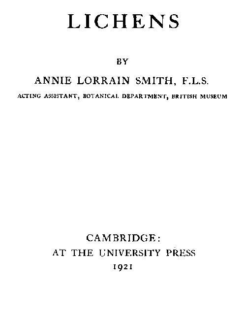
Fig. 99. Dermatocarpon miniatum Th. Fr. Vertical section of thallus and carpogonial group × 600 (after Baur).
b. Formation of Carpogonia. Stahl[607] had indicated that not only in gymnocarpous but also in angiocarpous lichens, it would be found that carpogonia were formed as in Collema. Baur[608] justified this surmise, and demonstrated the presence of ascogonia in groups of three to eight, with trichogynes that reached the surface in Endocarpon (Dermatocarpon) miniatum (Fig. 99). It is one of the few foliaceous Pyrenolichens, and the leathery thallus is attached to the substratum by a central point, thus allowing in the thallus not only peripheral but also intercalary growth, the latter specially active round the point of basal attachment; carpogonia may be found in any region where the tissue is newly formed, and at any season. The upper cortex is composed of short-celled thick-walled hyphae, with branching vertical to the surface, and so closely packed that there is an appearance of plectenchyma; the medullary hyphae are also thick-walled but with longer cells. The carpogonia of this species arise as a branch from the vegetative hyphae and are without special covering hyphae, so frequent a feature in other lichens. The trichogynes bore their way through the compact cortex and rise well above the surface. After they have disappeared—presumably after fertilization—the vegetative hyphae round and between the ascogonia become active and travel upwards slightly converging to a central point. The asci begin to grow out from the ascogenous hyphae of the base before the vertical filaments have quite pierced the cortex.
Pyrenula nitida has also been studied by Baur[609]. It is a very common species on smooth bark, with a thin crustaceous thallus immersed among the outer periderm cells. Unlike most other lichens, it forms carpogonia in spring only, from February to April. A primordial coil of hyphae lies at the base of the gonidial layer, and, before there is any appearance of carpogonia, a thick strand of hyphae is seen to be directed upwards, so that a definite form and direction is given to the perithecium at a very early stage. The ascogonial cells which are differentiated are extremely small, and, like those of all other species examined, are uninucleate. There are five to ten carpogonia in each primordium; the trichogynes grow up through the hyphal strand and emerge 5-10 µ above the surface. After their disappearance, a weft of ascogenous tissue is formed at the base, and, at the same time, the surrounding vegetative tissue takes part in the building up of a plectenchymatous wall of minute dark-coloured cells. Further development is rapid and occupies probably only a few weeks.
In many of the pyrenocarpous lichens—Verrucariae and others—the walls of the paraphyses dissolve in mucilage as the spores become mature, a character associated with spore ejection and dispersal. In some genera and species, as in Pyrenula, they remain intact.
D. Apogamous Reproduction
Though fertilization by an externally produced male nucleus has not been definitely proved there is probability that, in some instances, the fruit may be the product of sexual fusion. There are however a number of genera and species in which the development is apogamous so far as any external copulation is possible and the sporiferous tissue seems to be a purely vegetative product up to the stage of ascus formation.
In Phlyctis agelaea Krabbe[610] found abundant apothecia developing normally and not accompanied by spermogonia; in Phialopsis rubra studied also by him the primordium arises among the cells of the periderm on which the lichen grows, and he failed to find any trace of a sexual act. In his elaborate study of Gloeolichens Forssell[611] established the presence of carpogonia with trichogynes in two species—Pyrenopsis phaeococca and P. impolita, but without any appearance of fertilization; in all the others examined, the origin of the fruit was vegetative. Wainio[612] records a similar observation in a species of Pyrenopsis in which there was formed a spiral ascogonium and a trichogyne, but the latter never reached the surface.
Neubner[613] claimed to have proved a vegetative origin for the asci in the Caliciaceae; but he overlooked the presence of spermogonia and his conclusions are doubtful.
Fünfstück[614] found apogamous development in Peltigera (including Peltidea) and his results have never been disputed. The ascogonial cells are surrounded at an early stage by a weft of vegetative hyphae. No trichogynes are formed and spermogonia are absent or very rare in the genus, though pycnidia with macrospores occur occasionally.
Some recent work by Darbishire[615] on the genus supplies additional details. The apothecial primordium always originated near the growing margin of the thallus, where certain medullary hyphae were seen to swell up and stain more deeply than others. These at first were uninucleate, but the nuclei increased by division as the cells became larger, and in time there was formed a mass of closely interwoven cells full of cytoplasm. “No coiled carpogonia can be made out, but these darkly stained cells form part of a connected system of branching hyphae coming from the medulla further back.” Long unbranched multi-septate hyphae—evidently functionless trichogynes—travelled towards the cortex but gradually died off. Certain of the larger cells—the “ascogonia”—grew out as ascogenous hyphae into which the nuclei passed in pairs and finally gave rise to the asci.
These results tally well with those obtained by M. and Mme Moreau[616], though they make no mention of any trichogyne. They found that the terminal cells of the ascogenous hyphae were transformed into asci, and the two nuclei in these cells fused—the only fusion that took place. In Nephromium, one of the same family, the case for apogamy is not so clear; but Fünfstück found no trichogynes, and though spermogonia were present on the thallus, they were always somewhat imperfectly developed.
Sturgis[617] supplemented these results in his study of other lichens containing blue-green algae. In species of Heppia, Pannaria, Hydrothyria, Stictina and Ricasolia, he failed to find any evidence of fertilization by spermatia.
Solorina, also a member of Peltigeraceae, was added to the list of apogamous genera by Metzger[618] and his work was confirmed and amplified by Baur[619]: certain hyphae of the gonidial zone branch out into larger ascogonial cells which increase by active intercalary growth, by division and by branching, and so gradually give rise to the ascogenous hyphae and finally to the asci. Baur looked on this and other similar formations as instances of degeneration from the normal carpogonial type of development. Moreau[620] (Fernand and Mme) have also examined Solorina with much the same results: the paraphyses rise first from cells that have been produced by the gonidial hyphae; later, ascogenous hyphae are formed and spread horizontally at the base of the paraphyses, finally giving rise at their tips to the asci. Metzger[618] had further discovered that spermogonia were absent and trichogynes undeveloped in two very different crustaceous lichens, Acarospora (Lecanora) glaucocarpa and Verrucaria calciseda, the latter a pyrenocarpous species and, as the name implies, found only on limestone.
Krabbe[621] had noted the absence of any fertilization process in Gyrophora vellea. At a later date, Gyrophora cylindrica was made the subject of exact research by Lindau[622]. In that species the spermogonia (or pycnidia) are situated on the outer edge of the thallus lobes; a few millimetres nearer the centre appear the primordia of the apothecia, at first without any external indication of their presence. The initial coil which arises on the lower side of the gonidial zone consists of thickly wefted hyphae with short cells, slightly thicker than those of the thallus. It was difficult to establish their connection with the underlying medullary hyphae since these very soon change to a brown plectenchyma. From about the middle of the ascogonial coil there rises a bundle of parallel stoutish hyphae which traverse the gonidial zone and the cortex and slightly overtop the surface. They are genetically connected at the base with the more or less spirally coiled hyphae, and are similar to the trichogynes described in other lichens. Lindau did not find that they had any sexual significance, and ascribed to them the mechanical function of terebrators or borers. The correctness of his deductions has been disputed by various workers: Baur[619] looks on these “trichogynes” as the first paraphyses. The reproductive organs in Stereocaulon were examined by G. Wolff[623], and the absence of trichogynes was proved, though spermogonia were not wanting. She also failed to find any evidence of fertilization in Xanthoria parietina, in which lichen the ascogenous hyphae branch out from an ascogonium that does not form a trichogyne.
Rosendahl[624], as already stated, could find no trichogynes in Parmelia glabra. In Parmelia obscurata, on the contrary, Bitter[625] found that carpogonia with trichogynes were abundant and spermogonia very rare. In other species of the subgenus, Hypogymnia, he has pointed out that apothecia are either absent or occur but seldom, while spermogonia are numerous, and he concludes that the spermatia must function as spores or conidia. Baur[626] however does not accept that conclusion; he suggests as probable that the male organs persist longer in a functionless condition than do the apothecia.
Still more recently Nienburg[627] has described the ascogonium of Baeomyces sp. and also of Sphyridium byssoides (Baeomyces rufus) as reduced and probably degenerate. His results do not disprove those obtained by Krabbe[628] on the same lichen (Sphyridium fungiforme). The apothecia are terminal on short stalks in that species. When the stalk is about ·5mm. in height, sections through the tip show numerous primordia (12 to 15) ranged below the outer cortex, though only one, or at most three, develop further. These ascogonial groups are connected with each other by delicate filaments, and Nienburg concluded that they were secondary products from a primary group lower down in the tissue. Spirals were occasionally seen in what he considered to be the secondary ascogonia, but usually the fertile cells lie in loose uncoiled masses; isolated hyphae were observed to travel upwards from these cells, but they never emerged above the surface.
Usnea macrocarpa—if Schulte’s[629] work may be accepted—is also apogamous, though in Usnea barbata Nienburg[627] found trichogynes (Fig. 95) and the various developments that are taken as evidence of fertilization. Wainio[630] had demonstrated emergent straight trichogynes in Usnea laevis but without any sign of fertilization.
E. Discussion of Lichen Reproduction
In Ascolichens fertilization by the fusion of nuclei in the ascogonium is still a debated question. The female organ or carpogonium, as outlined above, comprises a twisted or spirally coiled multi-septate hypha, with a terminal branch regarded as a trichogyne which is also multi-septate, and through which the nucleus of the spermatium must travel to reach the female cell. It is instructive to compare the lichen carpogonium with that of other plants.
a. The Trichogyne. In the Florideae, or red seaweeds, in which the trichogyne was first described, that organ is merely a hair-like prolongation of the egg-cell and acts as a receptive tube. It contains granular protoplasm but no nucleus and terminates in a shiny tip covered with mucilage. The spermatium, unlike that of lichens, is a naked cell, and being non-motile is conveyed by water to the tip of the trichogyne to which it adheres; the intervening wall then breaks down and the male nucleus passes over. After this process of fertilization a plug of mucilage cuts off the trichogyne, and it withers away.
In Coleochaete, a genus of small freshwater green algae, a trichogyne is also present in some of the species: it is again a prolongation of an oogonial cell.
In the Ascomycetes, certain cells or cell-processes associated with the ascogonium have been described as trichogynes or receptive cells. In one of the simpler types, Monascus[631], the “trichogyne” is a cell cut off from the ascogonial cell. When fertilization takes place, the wall between the two cells breaks down to allow the passage of the male nucleus, but closes up when the process is effected. In Pyronema confluens[632] it is represented by a process from the ascogonial cell which fuses directly with the male cell. A more elaborate “trichogyne” has been evolved in Lachnea stercorea[633], another Discomycete: in that fungus it takes the form of a 3-5-septate hypha with a longer terminal cell; it rises from some part of the ascogonial cell but has no connection with any process of fertilization, so that the greater elaboration of form is in this case concomitant with loss of function.
In the Laboulbeniaceae, a numerous and very peculiar series of Ascomycetes that live on insects, there are, in nearly all of the reproductive bodies, a carpogonial cell, a trichophoric cell and a trichogyne. The last-named organ is in some genera a simple continuous cell, in others it is septate and branched, occasionally it is absent[634]. The male cells are spermatia of two kinds, exogenous or endogenous, and the plants are monoecious or dioecious. Laboulbeniaceae have no connection with lichens. Faull[635], a recent worker on the group, states that though he observed spermatia attached to the trichogynes, he was not able to demonstrate copulation (possibly owing to over-staining), nor could he trace any migration of the nucleus through the trichophoric cell down to the carpogonial cell. In two species of Laboulbenia that he examined there were no antheridia, and the egg-cell acquired its second nucleus from the neighbouring trichophoric cell. These conjugate nuclei divided simultaneously and the two daughter nuclei passed on to the ascus and fused, as in other Ascomycetes, to form the definitive nucleus.
Convincing evidence as to the importance of the trichogyne in fungi was supposed, until lately, to be afforded by the presence and functional activity of that organ associated with spermogonia in a few Pyrenomycetes—in Poronia, Gnomonia and Polystigma. Poronia was examined by M. Dawson[636] who found that a trichogyne-like filament distinct from the vegetative hyphae rose from the neighbourhood of the ascogonial cells. It took an upward course towards the exterior, but there was no indication that it was ever receptive. In Gnomonia erythrostoma and in Polystigma rubrum spermogonia with spermatia—presumably male organs—are produced in abundance shortly before the ascosporous fruit is developed. The spermatia in both cases exhibit the characters of male cells, i.e. very little cytoplasm and a comparatively large nucleus that occupies most of the cell cavity, along with complete incapacity to germinate. Brooks[637] found in Gnomonia that tufts of the so-called trichogynes originated near the ascogonial cells, but they were “mere continuations of ordinary vegetative hyphae belonging to the coil.” They are septate and reach the surface, and the tip-cell is longer than the others as in the lichen trichogyne.
A somewhat similar arrangement is present in Polystigma, in which Blackman and Welsford[638] have proved that the filaments, considered as trichogynes by previous workers, are merely vegetative hyphae. A trichogyne-like structure is also present in Capnodium, one of the more primitive Pyrenomycetes, but it has no sexual significance.
Lindau[639] in his paper on Gyrophora suggested that the trichogyne in lichens acted as a “terebrator” or boring apparatus, of service to the deeply immersed carpogonium in enabling it to reach the surface. Van Tieghem[640] explained its presence on physiological grounds as necessary for respiration, a view also favoured by Zukal[641], while Wainio[642] and Steiner[643] see in it only an “end-hypha,” the vigorous growth of which is due to its connection with the well-nourished cells of the ascogonium.
Lindau’s view has been rejected by succeeding writers: as has been already stated, it is the paraphyses that usually open the way outward for the apothecium. Van Tieghem’s theory has been considered more worthy of attention and both Dawson and Brooks incline to think that the projecting filaments described above may perform some service in respiration, even though primarily they may have functioned as sexual receptive organs.
There is very little support to be drawn from fungi for the theory that the presence of a trichogyne necessarily entails fertilization by spermatia. Lichens in this connection must be judged as a class apart.
It has perhaps been too lightly assumed that the trichogyne in lichens indicates some relationship with the Florideae[644]. Such a view might be possible if we could regard lichens and Florideae as derived from some common remote ancestor, though even then the difference in spore production—in one case exogenous, and in the other in asci and therefore endogenous—would be a strong argument against their affinity. But all the evidence goes to prove that lichens are late derivatives of fungi and have originated from them at different points. Fungi are interposed between lichens and any other ancestors, and inherited characters must have been transmitted through them. F. Bachmann’s suggestion[645] that Collema pulposum should be regarded “as a link between aquatic red algae and terrestrial ascomycetes such as Pyronema and the mildews” cannot therefore be accepted. It seems more probable that the lichen trichogyne is a new structure evolved in response to some physiological requirement—either sexual or metabolic—of the deeply embedded fruit primordium.
b. The Ascogonium. In fungi there is usually one cell forming the ascogonium, a coenogamete, which after fertilization produces ascogenous hyphae. There are exceptions, such as Cutting[646] found in Ascophanus carneus, in which it is composed of several cells in open contact by the formation of wide secondary pores in the cell-walls. In lichens the ascogonium is divided into a varying number of uninucleate cells. Darbishire[647] (in Physcia) and Baur[648] (in Anaptychia) have described an opening between the different cells, after presumed fertilization, that might perhaps constitute a coenogamete. Ascogenous hyphae arise from all, or nearly all the cells, whether fertilized by spermatia or not, and asci continue to be formed over a long period of time. There may even be regeneration of the entire fruiting body as described in Graphis elegans and in Pertusaria, apparently without renewed fertilization.
Spermogonia (or pycnidia) and the ascosporous fruits generally grow on the same thallus, though not unfrequently only one of the two kinds is present. As the spermogonia appear first, while the apothecia or perithecia are still in the initial stages, that sequence of development seems to add support to the view that their function is primarily sexual; but it is equally valid as a proof of their pycnidial nature since the corresponding bodies in fungi precede the more perfect ascosporous fruits in the life-cycle.
The differences in fertility between the two kinds of thallus in Collema crispum may be recalled[649]. Baur considered that development of the carpogonia was dependant on the presence of spermatia: a strong argument for the necessity of fertilization by these. The conditions in Parmelia acetabulum, also recorded by Baur, lend themselves less easily to any conclusion. On the thallus of that species the spermogonia and carpogonia present are out of all proportion to the very few apothecia that are ultimately formed. Though Baur suggested that cross-fertilization might be necessary, he admits that the development may be vegetative and so uninfluenced by the presence or absence of spermatia.
It is the very frequent occurrence of the trichogyne as an integral part of the carpogonium that constitutes the strongest argument for fertilization by spermatia. There is a possibility that such an organ may have been universal at one time both in fungi and in lichens, and that it has mostly degenerated through loss of function in the former, as it has disappeared in many instances in lichens. Again, there is but a scanty and vestigial record of spermogonia in Ascomycetes. They may have died out, or they may have developed into the asexual pycnidia which are associated with so many species. If we take that view we may trace the same tendency in lichens, as for instance in the capacity of various spermatia to germinate, though in lichen spermogonia there has been apparently less change from the more primitive condition. It is also possible that some process of nuclear fusion, or more probably of conjugation, takes place in the ascogonial cells, and that in the latter case the only fusion, as in some (or most) fungi, is between the two nuclei in the ascus.
If it be conceded that fully developed carpogonia with emergent trichogynes, accompanied by spermogonia and spermatia, represent fertilization, or the probability of fertilization, then the process may be assumed to take place in a fairly large and widely distributed series of lichens. Copulation between the spermatium and the trichogyne has been seen by Stahl[650], Baur[651] and by F. Bachmann[652] in Collema. In Physcia pulverulenta Darbishire[653] could not prove copulation in the earlier stages, but he found what he took to be the remains of emptied spermatia adhering to the tips of old trichogynes. Changes in the trichogyne following on presumed copulation have been demonstrated by several workers in the Collemaceae, and open communication as a result of fertilization between the cells of the ascogonium has been described in two species. This coenocytic condition of the ascogonium (or archicarp), considered by Darbishire and others as an evidence of fertilization, has been demonstrated by Fitzpatrick[654] in the fungus Rhizina undulata. The walls between the cells of the archicarp in that Ascomycete became more or less open, so that the ascogenous hyphae growing from the central cells were able easily to draw nutrition from the whole coenocyte, but no process of fertilization in Rhizina preceded the breaking down of the septa and no fusion of nuclei was observed until the stage of ascus formation.
The real distinction between fertile and vegetative hyphae lies, according to Harper[655], in the relative size of the nuclei. F. Bachmann speaks of one large nucleus in the spermatium of Collema pulposum which would indicate sexual function. There is however very little nuclear history of lichens known at any stage until the beginning of ascus formation, when fusion of two nuclei certainly take place as in fungi to form the definitive nucleus of the ascus.
The whole matter may be summed up in Fünfstück’s[656] statement that: “though research has proved as very probable that fertilization takes place, it is an undoubted fact that no one has observed any such process.”
F. Final Stages of Apothecial Development
The emergence of the lichen apothecium from the thallus, and the form it takes, are of special interest, as, though it is essentially fungal in structure, it is subject to various modifications entailed by symbiosis.
a. Open or closed Apothecia. Schwendener[657] drew attention to two types of apothecia directly influenced by the thallus: those that are closed at first and only open gradually, and those which are, as he says, open from the first. The former occur in genera and species in which the thallus has a stoutish cortex, as, for instance, in Lobaria where the young fructification has all the appearance of an opening perithecium. The open apothecia (primitus aperta) are found in non-corticate lichens, in which case the pioneer paraphyses arrive at the surface easily and without any converging growth. Similar apothecia are borne directly on the hypothallus at the periphery, or between the thalline areolae, and they are also characteristic of thin or slender thalli as in Coenogonium.
In both types of apothecium, the paraphyses pierce the cortex (Fig. 100) and secure the emergence of the developing ascomata.
Fig. 100. Physcia ciliaris DC. Vertical section of apothecium still covered by the cortex. a, paraphyses; b, hypothecium; c, gonidia of thallus and amphithecium. × 150 (after Baur).
b. Emergence of the Ascocarp. Hue[658] has taken up this subject in recent years and has described the process by which the vegetative hyphae surrounding the fruit primordium, excited to active growth by contact with the generative system, take part in the later stages of fruit formation. The primordium generally occupies a position near to, or just within, the upper medulla, and the hyphae in contact with it soon begin to branch freely in a vertical direction, surrounding the developing fruit and carrying it upwards generally to a superficial position. The different methods of the final emergence give two very distinct types of mature apothecium: the lecideine in which the gonidial zone takes no part in the upward growth, and the lecanorine into which the gonidia enter as an integral part.
In the lecideine series (Fig. 101) the encircling hyphae from the upper medulla rise as a compact column through the gonidial zone to the surface of the thallus; they then spread radially before curving up to form the outer wall or “proper margin” round the spore-bearing disc. The branching of the hyphae is fastigiate with compact shorter branches at the exterior. In such an apothecium gonidia are absent both below the hypothecium and in the margins.
Fig. 101. Lecidea parasema Ach. Vertical section of thallus and apothecium with proper margin only × ca. 50.
In lecanorine development the ascending hyphae from the medulla, in some cases, carry with them algal cells which multiply and spread as a second gonidial layer under the hypothecium (Fig. 102). These hyphae may also spread in a radial direction while still within the thallus and give rise to an “immersed” apothecium which is lecanorine as it encloses gonidia within its special tissues, for example, in Acarospora and Solorina. But in most cases the lecanorine fruit is superficial and not unfrequently it is raised on a short stalk (Usnea, etc.); both the primary gonidial zone of the thallus and the outer cortex are associated with the medullary column of hyphae from the first and grow up along with it, thus providing the outer part of the apothecium, an additional “thalline margin” continuous with the thallus itself. It is an advanced type of development peculiar to lichens, and it provides for fertility of long continuance which is in striking contrast with the fugitive ascocarps of the Discomycetes.
Fig. 102. Lecanora tartarea Ach. Vertical section of apothecium. a, hymenium; b, proper margin or parathecium; c, thalline margin or amphithecium. × 30 (after Reinke).
The distinction between lecideine and lecanorine apothecia is of great value in classification, but it is not always easily demonstrable; it is occasionally necessary to examine the early stages, as in the more advanced the thalline margin may be pushed aside by the turgid disc and become practically obliterated.
The “proper margin” reaches its highest development in the lecideine and graphideine types. It is less prominent or often almost entirely replaced when the thalline margin is superadded, except in genera such as Thelotrema and Diploschistes which have distinct “double margins.”
There is an unusual type of apothecium in the genus Gyrophora. The fruit is lecideine, the thalline gonidia taking no part in the development. The growth of the initial ascogenous tissue, according to Lindau[659], is constantly towards the periphery of the disc so that a weak spot arises in the centre which is promptly filled by a vigorous sterile growth of paraphyses. This process is repeated from new centres again and again, resulting in the irregularly concentric lines of sterile and fertile areas of the “gyrose” fruit (Fig. 103). The paraphyses soon become black at the tips. Asci are not formed until the ascogenous layer has acquired a certain degree of stability, and spores are accordingly present only in advanced stages of growth.
Fig. 103. Apothecial gyrose discs of Gyrophora cylindrica Ach. × 12 (after Lindau).
G. Lichen Asci and Spores
a. Historical. The presence of spores, as such, in the lichen fruit was first established by Hedwig[660] in Anaptychia (Physcia) ciliaris. He rightly judged the minute bodies to be the “semina” of the plant. In that species they are fairly large, measuring about 50µ, long and 24µ thick, and as they are very dark in colour when mature, they stand out conspicuously from the surrounding colourless tissue of the hymenium. Acharius[661] also took note of these “semina” and happily replaced the term by that of “spores.” They may be produced, he states, in a compact nucleus (Sphaerophoron), in a naked disc (Calicium), or they may be embedded in the disc (Opegrapha and Lecidea). Sprengel[662] opined that the spores—which he figures—were true seeds, though he allows that there had been no record of their development into new plants. Luyken[663] made a further contribution to the subject by dividing lichens into gymnocarpous and angiocarpous forms, according as the spores, enclosed in cells or vesicles (thecae), were borne in an open disc or in a closed perithecium.
In his Systema of lichen genera Eschweiler[664], some years later, described and figured the spores as “thecae” enclosed in cylindrical asci. Fée[665] in contemporary works gave special prominence to the colour and form of the spores in all the lichens dealt with.
b. Development of the Ascus. The first attempt to trace the origin and development of lichen asci and spores was made by Mohl[666]. He describes the mother-cell (the ascus) as filled at first with a clouded granular substance changing later into a definite number—usually eight—of simple or septate spores. Dangeard[667] included the lichens Borrera (Physcia) ciliaris and Endocarpon (Dermatocarpon) miniatam among the plants that he studied for ascus and spore development. He found that in lichens, as in fungi, the ascus arose usually from the penultimate cell of a crooked hypha (Fig. 104) and that it contained at first two nuclei derived from adjoining cells. These nuclei are similar in size to those of the vegetative hyphae, and in each there is a large nucleolus with chromatin material massed on one side. Fusion takes place, as in fungi, between the two nuclei, and the secondary or definitive nucleus thus formed divides successively to form the eight spore-nuclei. Baur[668] and Nienburg[669] have confirmed Dangeard’s results as regards lichens, and René Maire[670] has also contributed important cytological details on the development of the spores. In Anaptychia (Physcia) ciliaris he found that the fused nucleus became larger and that a synapsis stage supervened during which the long slender chromatin filaments became paired, and at the same time shorter and thicker. The nuclear membrane disappeared as the chromatin filaments were united in masses joined together by linin threads which also disappeared later. At the most advanced stage observed by Maire there was visible a nucleolus embedded in a condensed plasma and surrounded by eight medianly constricted filaments destined to form the equatorial plate. A few isolated observations were also made on the cytology of the ascus in Peltigera canina, in which lichen the preceding ascogonial development is wholly vegetative. The secondary nucleus was seen to contain a chromatin mass and a large nucleolus; in addition two angular bodies of uncertain signification were associated with the nucleolus, each with a central vacuole. The nucleolus disappeared in the prophase of the first division and four double chromosomes were then plainly visible. The succeeding phases of the first and the second nuclear division were not seen, but in the prophase of the third it was possible to distinguish four chromatin masses outside the nucleolus. The slow growth of the lichen plant renders continuous observation extremely difficult.
Fig. 104. Developing asci of Physcia ciliaris DC. × 800 (after Baur).
F. Bachmann[671] was able to make important cytological observations in her study of Collema pulposum. As regards the vegetative and ascogonial nuclei, five or perhaps six chromosomes appeared on the spindle when the nucleus divided. In the asci, the usual paired nuclei were present in the early stages and did not fuse until the ascus had elongated considerably. After fusion the definitive nucleus enlarged with the growth of the ascus and did not divide until the ascus had attained full size. The nucleolus was large, and usually excentric, and there were at first a number of chromatin masses on an irregular spirem. In synapsis the spirem was drawn into a compact mass, but after synapsis, “the chromatin is again in the form of a knotty spirem.” In late prophases the chromosomes, small ovoid bodies, were scattered on the spindle; later they were aggregated in the centre, and, in the early metaphase, about twelve were counted now split longitudinally. There were thus twice as many chromosomes in the first division in the ascus as in nuclear divisions of the vegetative hyphae. F. Bachmann failed to see the second division; there were at least five chromosomes in the third division.
Considerable importance is given to the number of the chromosomes in the successive divisions in the ascus since they are considered to be proof of a previous double fusion—in the ascogonium and again in the ascus—necessitating, therefore, a double reduction division to arrive at the gametophytic or vegetative number of five or six chromosomes in the third division in the ascus. There have been too few observations to draw any general conclusions.
c. Development of Spores. The spore wall begins to form, as in Ascomycetes, at the apex of the nucleus with the curving over of the astral threads, the nucleus at that stage presenting the figure of a flask the neck of which is occupied by the centrosome. The final spore-nucleus, as observed by Maire, divides once again in Anaptychia and division is followed by the formation of a median septum, the mature spore being two-celled. In Peltigera the spore is at first ovoid, but both nucleus and spore gradually elongate. The fully formed spore is narrowly fusiform and by repeated nuclear division and subsequent cross-septation it becomes 4- or even 5-6-celled.
The spores of lichens are wholly fungoid, and, in many cases, form a parallel series with the spores of the Ascomycetes. Markings of the epispore, such as reticulations, spines, etc., are rarely present (Solorina spongiosa), though thickening of the wall occurs in many species (Pertusariae, etc.), a peculiarity which was first pointed out by Mohl[672] who contrasted the spore walls with the delicate membranes of other lichen cells. Some spores, described as “halonate,” have an outer gelatinous covering which probably prevents the spore from drying up and thus prolongs the period of possible germination. Both asci and spores are, as a rule, more sparingly produced than in fungi; in many instances some or all of the spores in the ascus are imperfectly formed, and the full complement is frequently lacking, possibly owing to some occurrence of adverse conditions during the long slow development of the apothecium. In the larger number of genera and species the spores are small bodies, but in some, as for instance in the Pertusariae and in some Pyrenocarpeae, they exceed in size all known fungus spores. In Varicellaria microsticta, a rare crustaceous lichen of high mountains, the solitary 1-septate spore measures up to 350 µ, in length and 115 µ in width. Most spores contain reserve material in the form of fat, etc., many are dark-coloured; Zukal[673] has suggested that the colour may be protective.
Their ejection from the ascus at maturity is caused by the twofold pressure of the paraphyses and the marginal hyphae on the addition of moisture. The spores may be shot up at least 1 cm. from the disc[674].
d. Spore Germination. Meyer[675] was the first who cultivated lichen spores and the dendritic formation which he obtained by growing them on a smooth surface was undoubtedly the prothallus (or hypothallus) of the lichen. Actual germination was however not observed till Holle[676] in 1846 watched and figured the process as it occurs in Physcia ciliaris.
Spores divided by transverse septa into two or more cells, as well as those that have become “muriform” by transverse and longitudinal septation, may germinate from each cell.